

NUR 265 EXAM 2 - Exam 2
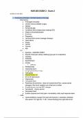
EXAM 2 NUR 265 ● Pulmonary embolism: clot that travels to the lungs ○ Risk factors ■ Prolonged immobility ■ Central venous catheter surgery ■ Obesity ■ Advancing age ■ Conditions that increase blood clotting (DIC) ■ Distort of thromboembolism ■ Smoking ■ Pregnancy ■ Hormonal birth control (estrogen therapy) ■ Heart failure ■ Stroke ■ Cancer ■ Trauma ■ Afib ○ S/s: ■ Dyspnea - SUDDEN ONSET ■ Pleuritic chest pain (sharp, stabbing type pain on inspiration) ■ Crackles ■ Wheezes ■ Apprehension ■ Anxiety ● Give O2 ■ Restlessness ■ Impending doom ■ Cough (productive or dry) ■ Tachypnea ■ Pleural friction rub ■ S3 or s4 heart sound ■ Diaphoresis ■ Low grade fever ■ Petechiae (fat embolism , does not impede blood flow, causes actual damage to the blood vessels) over chest and a axillae ■ If really big you can see EKG changes ■ hemoptysis - bloody sputum ■ Decreased Sao2 ■ Sudden dyspnea and chest pain= immediately notify rapid response team ○ Labs: ■ Hyperventilation (caused from pain and hypoxia) = respiratory alkalosis (low paco2 35, high PH 7.45) = blood shunting from right side to left lOMoARcPSD|8945469 ○ Dx: side without picking up O2 from the = respiratory acidosis (high paco2 45, low PH 7.35)= build up of lactic acid = metabolic acidosis (low HCO3 22, low PH 7.35) ■ D-dimer rises (positive) ■ Pulmonary angiography = gold standard ● Only if stable ● Inject dye, use imaging ■ CT ■ Chest X-ray ■ Doppler ultrasound ○ Nursing intervention: ■ Call rapid ■ O2 - use pulse ox ● Nasal cannula ● Mask ● Mechanical ventilation ■ Tele ■ IV access ■ Monitor VS. lung sounds and cardiac/ respiratory status Q1-2hrs ● Assess for and document increasing dyspnea, dysrhythmias, JVD, pedal or sacral edema, crackles, cyanosis ■ CTPA, pulmonary angiography ■ Bleeding precautions ● Monitor and record amount of bleeding ● Asses Q2 hours ■ Measure abdominal girth Q8 Hours ■ Monitor labs daily ● Monitor CBC to watch for blood loss ○ Blood loss= RBC, plasma ○ Monitor platelet count = decreased platelet count = HIIT ■ Drug therapy ■ Make sure antidote is on the floor ● Anticoagulants - keep clots from getting bigger ○ Unfractionated heparin ■ Check PTT (normal 20-30) before administering (range between 1.5-2.5 times the control) (therapeutic 46-70) (75 = complication) ■ 5-10 days (for 24 hours) ■ Protamine sulfate = antidote ○ Then transferred to oral warfarin ■ Monitor INR (2.0-3.0) ■ Vitamin K = antidote lOMoARcPSD|8945469 ○ Heparin 5-10 days, most patients started on warfarin on day 1-2, both are continued together until the INR reaches 2-3, heparin will be continued for 24 hours after INR is 2 ● ○ Heparin induced thrombocytopenia ■ Body creates antibodies to the heparin = increased thrombin - prothrombin = increased clotting ■ Risk factors include: ● Duration of heparin use longer than 1 week ● Exposure to unfractionated heparin ● Post surgical prothrombin prophylaxis ● Being female ■ S/s: ● DVT ● PE ● Thrombocytopenia (hallmark sign) = platelets 150,000 ● Can through thrombus ■ Treatment: ● Argatroban and lepirudin (direct thrombin inhibitors) ● Inferior vena cava filtration - bedside procedure ○ Filter that catches things before it gets to the lungs ● Mechanical ventilation for respiratory acidosis + pao2 60 = respiratory failure ● ● TPA ○ Antidote = clotting factors, frozen plasma, aminocaproic acid ● Mechanical ventilation ○ Mode: ■ A/C : assistive control ventilation ● Most restrictive - vent takes control of breathing completely ● Complications: ○ Hyperventilation ○ Respiratory alkalosis ( paco2 35, ph 7.45) ● Resting mode: to try to allow patient to breath on their own, if not working then the vent takes over and breathing pattern is established ■ SIMV: synchronized intermittent mandatory ventilation ● Can be used as a main ventilator or as a weaning method ● Allows for spontaneous breathing at patients own rate ■ CPAP: continuous positive airway pressure ○ Rate lOMoARcPSD|8945469 ○ Fractionated inspired O2 (FiO2) ○ Tidal volume (Vt) ○ Positive end expirations pressure (PEEP) ■ Keep alveoli open longer ■ Complications: ● Pneumothorax ● Decreased pre load ○ Mechanical ventilator care ■ Lung assessment ■ Frequent VS ■ Pulmonary hygiene ■ Monitor ABG ■ ETT cuff/ pilot balloon ■ NPO ■ Skin care ■ I&O / Foley catheter ■ VTE prophylaxis ○ Pneumonia ■ Remove water from circuits ■ HOB up 30 degrees ■ Turn Q2hrs ■ Suction ■ Oral care ■ Lip moisturizer ■ Percussion ○ High pressure alarms ■ Blocked airway ■ Biting tube ■ Kinks ■ Coughing ■ Tension pneumothorax ■ Pulmonary edema ■ Psychomotor agitation ■ Pain ○ Low pressure alarms ■ Not getting enough air in or out ■ Air leak in the cuff ■ Disconnect tubing ■ Patient not breathing ● Acute respiratory distress syndrome (ARDS): stiffness= things in alveoli= refractory hypoxemia ○ Causes respiratory tissue to become inflamed which becomes stiff and then allows debri and other things into the alveoli - things that should not be in pockets lOMoARcPSD|8945469 ■ Gas exchange happens in the alveoli ■ Acute in onset, results from lung injury ○ Subtle changes in early stages (s/s) ■ Refractory hypoxemia : when a pt is given O2 and they remain hypoxemia - 100% O2 and still hypoxia = ARDS ■ Decreased PAO2 (80-100) ■ Hyperpnea ● Increased work effect ● Noisy respiration ■ Hypoxia ■ Cyanosis ■ Pallor ■ Intercostal and substernal retractions ■ Diaphoresis ■ Clear lung sounds early - abnormal lung sounds are not heart on auscultation because the edema occurs first in the interstitial spaces not in the airways ● Late signs = crackles ● Dysrhythmias ○ Common causes: ■ Shock ■ Trauma ■ Pancreatitis ■ Fat/ amniotic fluid emboli ■ Sepsis ■ Aspiration - especially stomach contents ■ Pneumonia ■ Inhaling toxic fumes ■ Drug ingestion (heroine, opioids, aspirin) ■ Hemolytic disorders ■ Cardiopulmonary bypass ■ Multiple blood transfusions ● Usually within 6 hours of transfusion ■ Water aspiration from submersion ● Especially fresh water ■ COVID ■ Nervous system injury ■ Fat embolism ■ Amniotic fluid emboli ○ Requires immediate intervention ○ Difficult to diagnose ■ ABG ● Low PAO2 ● Sputum cultures lOMoARcPSD|8945469 ● Chest X-ray ○ Will show ground glass appearance “whited out” ● Hemodynamic monitoring is ****** waiting for an answer! ○ PAP and PCWP ○ May lead to complications ■ SIRS ■ Multi organ failure ■ Respiratory failure - respiratory acidosis (ph 7.28, Co2 55) and a PAO2 of 60, respiratory rate goes up, oxygen saturation goes down ○ ARDS management ■ Supportive care ■ On vent ● Will be flipped prone to relieve the pressure off the lungs ● 100% FIO2 ● PEEP ○ Assess for TP (tension pneumothorax ) ○ Started at 5 cm H2O and increased to keep oxygen saturation adequate ○ Low tidal volume (6 ml/kg of body weight) have shown to prevent lung injury ○ Prone position ○ Tracheostomy is considered if artificially airway is needed for longer than 10-14 days ● APRV - does not allow for spontaneous breathing between mandatory breaths ● HFOV- sedation and paralytics used especially with this ■ Antibiotics ■ Sedation vacation ■ Position and turning ■ Nutritional support ● Tube feeding / TPN ■ Corticosteroids ■ Modify the inflammatory response and reduce oxidative stress are older investigation ● Vitamins C and E, N-acetylcysteine, nitric oxide, surfactant replacement ■ Minimal fluids - conservative fluid therapy to improve lung function and shorten duration on vent ■ Diuretics to maintain fluid balance ■ Usually after 14 days they are either dead or have chronic disease with neuropsychological deficits ● Pleural effusion : buildup of fluid in the pleural space ○ Causes: lOMoARcPSD|8945469 ■ Heart failure ■ Kidney failure ■ Infection ■ Malignancy ■ Cirrhosis ○ Symptoms: ■ Every time I take a breath it hurts - chest pain ■ Asymmetrical chest expansion ■ Difficulty breathing ● Hurts more when they breath ■ Dry or productive cough ■ Fever - infection ■ Pleasurable friction rub ■ Egophony - when you put stethoscope on their back on their skin and they say the letter “E” it will sound like “E” on normal lung but sounds like “A” on pleural effusion ■ Decreased tactile Fremitus - use hands to go down bilateral posterior lungs on their skin, they say “99”, when hand is on pleasurable effusion fluid the vibration on the hands will be decreased ○ Diagnosis ■ Chest X-ray ■ CT scan ○ Treatment : ■ Antibiotics ■ Steroids ■ Thoracentesis ■ Diuretics ● Chest traumas ○ Rib fractures ■ Blunt trauma to the chest ■ Risk for : ● Pulmonary contusion ● Pneumothorax ● Hemothorax ■ S/s: ● Pain with movement ■ Tx: ● Treat the pain ● Usually no other treatment needed ● Don’t wrap or bind ● Suction and incentive spirometer bedside ○ Pulmonary contusion: giant bruise on the ribs ■ Very common in blunt thoracic trauma, like a giant bruise on the lungs lOMoARcPSD|8945469 ■ Complications ● Potentially lethal ● Respiratory failure developers over time hemorrhaging and edema occur in the lungs ■ S/s: ● Hypoxia and dyspnea ● Hemolysis ● Crackles, wheezing, decreased sounds in the area ■ Dx: ■ Tx ● Chest X-ray ● Usually placed on a mechanical vent if contusion gets bigger and starts to go into respiratory failure ● Don’t turn them onto the side that is injured, good lung down ● IVF, O2, PEEP ○ Monitor for tension pneumothorax ● Avoid analgesics that cause respiratory depression ● Intercostal nerve block for pain if severe ■ Complications: ● ARDS ● Tension pneumothorax ● Death ● Moderate flowers ○ Flail chest ■ Can involve one side or both sides, blunt force trauma, incorrect chest compressions ■ Paradoxical chest movement ● Chest move in for inspiration ● Chest moves out for expiration ● A group of ribs that move like a seesaw - goes opposite of the movement that should be occurring with breathing ● Dyspnea ● Cyanosis ● Tachycardia ● Hypotension ■ Tx: ● May require mechanical vent ○ Hypoxemia and hypercapnia ● Give IV fluids ● Manage hypotension / shock - watch for s/s: ○ Decreased BP, increased HR ● Continuous O2 ● ABGs ● Tele lOMoARcPSD|8945469 ● Surgery - cage ● Pain management ○ Analgesic, epidural, nerve bock ○ Pneumothorax: air trapped in pleural space, uneven expansion ■ Air in the pleural space - usually higher ■ Typically results from blunt chest trauma ■ S/s: ● Absent / decreased breath sounds ● Asymmetrical lung expansion ● Pain ● Tachypnea ● Subcutaneous emphysema - crepitus (rice crispies) - every time they exhale the air moves out into the space ● Hyper resonance on percussion ■ Tx: ● Chest tube with suction to suck the air out ○ Hemothorax: blood in pleasurable space ■ Blunt chest trauma - blood in the pleural space - usually lower ■ S/s: ● Dyspnea ● Tachypnea ● Pain ● Absent breath sounds ■ Tx: ● Chest tube ○ Watch for abrupt cessation (clot) ■ 70 ml/hours = call dr ■ If drainage stops out of nowhere = call dr ■ An open thoracotomy is needed when there is initial blood loss of 1000 ml from the chest or persistent bleeding at the rebate of 150-200 ml/hr over 3-4 hours ■ Blood lost can be infused back into the patient after processing if needed ○ Should taper as it resolves ○ Shouldn’t she’s tidaling in water seal chamber - patient breathing ○ Do not want to see continuous bubbles in the air leak monitor ○ Milking the tube = good, hand over hand ○ Stripping the tube = bad - don’t compress the tube ● Mechanical ventilation ■ Nursing interventions lOMoARcPSD|8945469 ○ Tension pneumothorax: air in pleural space that can’t move out, tracheal deviation ■ Emergency ■ Air leak in the lung field - air can’t move in and out ■ S/s: ● Tracheal deviation ● Absent breath sounds ● Cyanosis ● Distended neck veins ● Dyspnea ■ Tx: ● Chest tube first ○ Water in the water seal chamber sodium normal rises 2 - 4 inches during inhalation and falls during exhalation - called tidaling ○ When to call the MD ■ Tracheal deviation: tension pneumothorax s/s ■ Sudden onset or increase in dyspnea ■ O2 stat less than 90% ■ Visible eyelets on chest tube ■ Tube falls out of chest = 1st cover area with dry gauze ■ Tube disconnects from drainage system = 1st place in sterile water ■ Drainage stops within 1st 24 hours ■ Bubbling occurs continuously in the water seal chamber - indicates an air leak ■ Drainage fluid must never fill to the point that it comes into contact with any tubes, if the tubing from the patient meters the fluid, drainage stops and can lead to a tension pneumothorax ● Then mechanical vent ● Complex endocrine / metabolism - anterior pituitary ● Thyroid: produces hormone ○ T3 &T4 increase O2 use and heat production. Stimulates HR and stroke volume ○ Calcitonin : regulates serum calcium levels ○ When anterior pituitary and hypothalamus gland senses low levels of hormones, sends thyroid stimulating hormone to thyroid to produce more T3 and T4 ○ Hyperthyroidism: over active metabolism, high T3, T4, low TSH ■ Causes: ● Grave’s disease ● Autoimmune (Hashimoto’s thyroiditis)
Document information
- Uploaded on
- May 6, 2025
- Number of pages
- 26
- Written in
- 2024/2025
- Type
- Exam (elaborations)
- Contains
- Questions & answers


