Bio Unit 3 Review
M4-1 Notes
• Ach binds to nicotinic receptors to contract
o Nicotinic for muscle contraction
o Nicotinic cholinergic = chemically-gated Na+ channel
• Motor end plate high concentration of Ach receptors
• Contraction happens as long as Ca2+ is inside cytoplasm
• Muscle twitch = 1 cycle of contraction and relaxation
• ATP needed for both contraction and relaxation
• During E-C coupling, skeletal muscle AP down t-tubules causes release of Ca2+ from SR
• DHP voltage-sensitive, so opens RyR gate with AP
• RyR is on SR
o RyR receptor is Ca2+ channel
• Ca2+ concentrations
o Very concentrated in SR
o High in ECM
o Low in sarcoplasm
o Low in axon terminus
• ATP moves myosin to cocked position via hydrolysis, releasing phosphate
• Anticholinesterase blocks acetylcholinesterase = increases amount of Ach in synapse
• Plasmapheresis filters antibodies out of blood
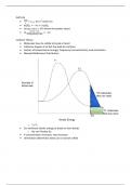
• Slow-twitch have a lot of myoglobin because need a lot of O2
o Myoglobin in muscle binds to O2
o Hemoglobin carries O2 in blood
• Fast-twitch have lighter color because less O2
o Quick, like eye-movements
• Smooth muscle criss-crossing myofilaments
o Muscle fibers in different directions within organs
• Skeletal muscle contraction
o Somatic motor neuron AP arrives at motor end plate
o Release of Ach from somatic motor neuron causes excitation of skeletal muscle
cell
o AP in skeletal muscle triggers Ca2+ signaling to contract sarcomeres
o Relaxation of muscle after contraction
, • First AP of somatic motor neuron à ACh release
• Second AP of skeletal muscle à sarcomere contraction
o Na+ entry = initial depolarization =skeletal muscle AP
o Perpetuated by VGSC down entire t-tubule
o Finishes long before muscle twitch
• Short refractory period
• Varicosities synthesize, store, and release neurocrines
• Smooth muscle needs more points of contact/more varicosities because longer
• Adrenergic in response to (nor)epinephrine
• Muscarinic in response to acetylcholine
• Ca2+ enters cytoplasm
o From outside cell by VGCC
o From SR by Ca2+ binding to RyR on SR (calcium induced calcium release)
o From SR by IP3 binding to receptor and releasing calcium
• Calcium binds to calmodulin
o Calmodulin-Ca2+ activates myosin light-chain kinase
o Kinase adds phosphate group to myosin light chain = tighter binding
o Increases cross bridge formation = more contraction
• cAMP blocks calmodulin-Ca2+ complex
o In response to metabotropic signaling
o Prevents actomyosin binding = prevents phosphorylation = relaxation
• Tonic: sphincter, blood vessels
• Phasic: esophagus, bladder
M4-2 Notes
• Cardiovascular system needed to deliver O2 in/CO2 out quickly
• Heart on ventral side, between lungs
o Apex points left/inferior
o Base superior
• Pericardial sac encloses heart
o Epicardium and parietal pericardium enclose pericardial cavity
o Pericardial fluid prevents friction
• Epithelial endocardium lines luminal side
o Epicardium lines outer side
• Epicardium innermost layer of pericardial sac
• Afferent vesicles empty into atria
, • Atria = upper; ventricles = lower
• Efferent vesicles from ventricles
• Coronary arteries/veins circulation for heart muscle
• Deox blood to heart, oxy away
• Closed loops by pulmonary/systemic circulatory systems
• Tendons attach flaps to heart wall
o Chordae tendinae are connective tissue
o Connect to papillary muscles
• Flap = cuspid
o Tendons = chordae tendinae
o Muscles = papillary
• Papillary muscles attachment only, not open or close
• Semilunar valves 3 leaflets each, moon-shaped
o No chordae tendinae bc small
• Systole is contraction
• Diastole is relaxation
• Atria contract separately from ventricles
o But L&R atria contract together
o And L&R ventricles
• Blood pressure causes opening/closing of flaps
• Each heart beat pumps abt half of volume if relaxed
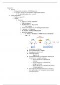
• Pacemaker potential depolarizes as reaches threshold = triggers AP
o If channels close at threshold; VGCC open at threshold
• Na+ in, Ca2+ in, K+ out
• Rate of depolarization = pace of autorhythmic
• Opening of VGCC = depolarization
• (Para)sympathetic controls rate of depolarization
• Contractile cell AP phases
o Depolarization: from Na+ influx via gap junction
o Initial repolarization: K+ efflux
o Plateau phase: Ca2+ influx, decreased K+ efflux
o Rapid repolarization: increased K+ efflux, decreased Ca2+ influx
• Plateau caused by Ca2+ influx lengthens AP
M4-1 Notes
• Ach binds to nicotinic receptors to contract
o Nicotinic for muscle contraction
o Nicotinic cholinergic = chemically-gated Na+ channel
• Motor end plate high concentration of Ach receptors
• Contraction happens as long as Ca2+ is inside cytoplasm
• Muscle twitch = 1 cycle of contraction and relaxation
• ATP needed for both contraction and relaxation
• During E-C coupling, skeletal muscle AP down t-tubules causes release of Ca2+ from SR
• DHP voltage-sensitive, so opens RyR gate with AP
• RyR is on SR
o RyR receptor is Ca2+ channel
• Ca2+ concentrations
o Very concentrated in SR
o High in ECM
o Low in sarcoplasm
o Low in axon terminus
• ATP moves myosin to cocked position via hydrolysis, releasing phosphate
• Anticholinesterase blocks acetylcholinesterase = increases amount of Ach in synapse
• Plasmapheresis filters antibodies out of blood
• Slow-twitch have a lot of myoglobin because need a lot of O2
o Myoglobin in muscle binds to O2
o Hemoglobin carries O2 in blood
• Fast-twitch have lighter color because less O2
o Quick, like eye-movements
• Smooth muscle criss-crossing myofilaments
o Muscle fibers in different directions within organs
• Skeletal muscle contraction
o Somatic motor neuron AP arrives at motor end plate
o Release of Ach from somatic motor neuron causes excitation of skeletal muscle
cell
o AP in skeletal muscle triggers Ca2+ signaling to contract sarcomeres
o Relaxation of muscle after contraction
, • First AP of somatic motor neuron à ACh release
• Second AP of skeletal muscle à sarcomere contraction
o Na+ entry = initial depolarization =skeletal muscle AP
o Perpetuated by VGSC down entire t-tubule
o Finishes long before muscle twitch
• Short refractory period
• Varicosities synthesize, store, and release neurocrines
• Smooth muscle needs more points of contact/more varicosities because longer
• Adrenergic in response to (nor)epinephrine
• Muscarinic in response to acetylcholine
• Ca2+ enters cytoplasm
o From outside cell by VGCC
o From SR by Ca2+ binding to RyR on SR (calcium induced calcium release)
o From SR by IP3 binding to receptor and releasing calcium
• Calcium binds to calmodulin
o Calmodulin-Ca2+ activates myosin light-chain kinase
o Kinase adds phosphate group to myosin light chain = tighter binding
o Increases cross bridge formation = more contraction
• cAMP blocks calmodulin-Ca2+ complex
o In response to metabotropic signaling
o Prevents actomyosin binding = prevents phosphorylation = relaxation
• Tonic: sphincter, blood vessels
• Phasic: esophagus, bladder
M4-2 Notes
• Cardiovascular system needed to deliver O2 in/CO2 out quickly
• Heart on ventral side, between lungs
o Apex points left/inferior
o Base superior
• Pericardial sac encloses heart
o Epicardium and parietal pericardium enclose pericardial cavity
o Pericardial fluid prevents friction
• Epithelial endocardium lines luminal side
o Epicardium lines outer side
• Epicardium innermost layer of pericardial sac
• Afferent vesicles empty into atria
, • Atria = upper; ventricles = lower
• Efferent vesicles from ventricles
• Coronary arteries/veins circulation for heart muscle
• Deox blood to heart, oxy away
• Closed loops by pulmonary/systemic circulatory systems
• Tendons attach flaps to heart wall
o Chordae tendinae are connective tissue
o Connect to papillary muscles
• Flap = cuspid
o Tendons = chordae tendinae
o Muscles = papillary
• Papillary muscles attachment only, not open or close
• Semilunar valves 3 leaflets each, moon-shaped
o No chordae tendinae bc small
• Systole is contraction
• Diastole is relaxation
• Atria contract separately from ventricles
o But L&R atria contract together
o And L&R ventricles
• Blood pressure causes opening/closing of flaps
• Each heart beat pumps abt half of volume if relaxed
• Pacemaker potential depolarizes as reaches threshold = triggers AP
o If channels close at threshold; VGCC open at threshold
• Na+ in, Ca2+ in, K+ out
• Rate of depolarization = pace of autorhythmic
• Opening of VGCC = depolarization
• (Para)sympathetic controls rate of depolarization
• Contractile cell AP phases
o Depolarization: from Na+ influx via gap junction
o Initial repolarization: K+ efflux
o Plateau phase: Ca2+ influx, decreased K+ efflux
o Rapid repolarization: increased K+ efflux, decreased Ca2+ influx
• Plateau caused by Ca2+ influx lengthens AP