Chapter 5: The Integumentary System
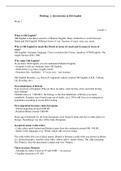
Functional Anatomy of the Skin
Two major components
1. Cutaneous membrane
2. Accessory structures
1. Cutaneous membrane
o Epidermis (epi, above)
Composed of stratified squamous epithelium
o Dermis
Papillary layer (areolar connective tissue)
Reticular layer (dense irregular connective tissue)
2. Accessory structures
o Hairs
o Nails
o Exocrine glands
Sebaceous glands
Sweat glands
o Sensory receptors and nerve fibers
o Arrector pili muscles
o Cutaneous plexus (network of blood vessels)
Functions of the integumentary system
Protection
Excretion
Body temperature regulation
Production of melanin and keratin
Vitamin D3 synthesis
Lipid storage
Sensory input
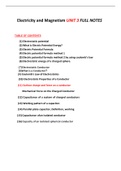
Epidermis overview
Multiple layers of cells (strata)
Primary cell type in epidermis is keratinocyte
o Body’s most abundant epithelial cell
o Continuously produced by stem cell division in deepest layers
o Shed at exposed surfaces
Deeper layers of epidermis form epidermal ridges
o Adjacent to dermal papillae (papilla, nipple-shaped mound)
o Increase surface area for better attachment
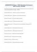
Fingerprints
© 2015 Pearson Education, Inc.
, Pattern of epidermal ridges on surface of fingertips
Determined by genes and intrauterine environment during fetal development
Unique pattern that does not change during lifetime
o Prints of these patterns (fingerprints) used to identify individual
“Thin” and “thick” refers to thickness of epidermis
Thin skin
o Covers most of body surface
o Contains four strata (layers)
o About as thick as a plastic sandwich bag (~0.08 mm)
Thick skin
o Found on palms of hands and soles of feet
o Contains five strata (layers)
o About as thick as a paper towel (~0.5 mm)
Epidermal layers overview
Entire epidermis lacks blood vessels
o Cells get oxygen and nutrients from capillaries in the dermis
o Cells with highest metabolic demand are closest to the dermis
o Takes about 7–10 days for cells to move from the deepest stratum to
the most superficial layer
o Cells in surface layer (stratum corneum) remain about 2 weeks before
being shed or washed away
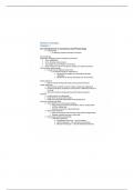
Epidermal layers deep to superficial
1. Stratum basale
o Basal (bottom) layer of the epidermis
o Attached to basement membrane by hemidesmosomes
o Most cells here are basal cells, stem cells that divide to replace more
superficial keratinocytes
o Merkel cells that respond to touch are also found here
2. Stratum spinosum (“spiny layer”)
o Composed of 8–10 layers of keratinocytes bound together by
desmosomes
Only looks spiny when on a prepared slide
Contains dendritic (Langerhans) cells
o Part of immune response defending against
microorganisms and superficial skin cancers
3. Stratum granulosum (“grainy layer”)
o Composed of 3–5 layers of keratinocytes
o Most cells have stopped dividing and started producing keratin and
keratohyalin
o Cells grow thinner and flatter
© 2015 Pearson Education, Inc.
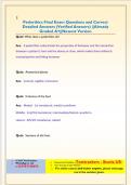
Functional Anatomy of the Skin
Two major components
1. Cutaneous membrane
2. Accessory structures
1. Cutaneous membrane
o Epidermis (epi, above)
Composed of stratified squamous epithelium
o Dermis
Papillary layer (areolar connective tissue)
Reticular layer (dense irregular connective tissue)
2. Accessory structures
o Hairs
o Nails
o Exocrine glands
Sebaceous glands
Sweat glands
o Sensory receptors and nerve fibers
o Arrector pili muscles
o Cutaneous plexus (network of blood vessels)
Functions of the integumentary system
Protection
Excretion
Body temperature regulation
Production of melanin and keratin
Vitamin D3 synthesis
Lipid storage
Sensory input
Epidermis overview
Multiple layers of cells (strata)
Primary cell type in epidermis is keratinocyte
o Body’s most abundant epithelial cell
o Continuously produced by stem cell division in deepest layers
o Shed at exposed surfaces
Deeper layers of epidermis form epidermal ridges
o Adjacent to dermal papillae (papilla, nipple-shaped mound)
o Increase surface area for better attachment
Fingerprints
© 2015 Pearson Education, Inc.
, Pattern of epidermal ridges on surface of fingertips
Determined by genes and intrauterine environment during fetal development
Unique pattern that does not change during lifetime
o Prints of these patterns (fingerprints) used to identify individual
“Thin” and “thick” refers to thickness of epidermis
Thin skin
o Covers most of body surface
o Contains four strata (layers)
o About as thick as a plastic sandwich bag (~0.08 mm)
Thick skin
o Found on palms of hands and soles of feet
o Contains five strata (layers)
o About as thick as a paper towel (~0.5 mm)
Epidermal layers overview
Entire epidermis lacks blood vessels
o Cells get oxygen and nutrients from capillaries in the dermis
o Cells with highest metabolic demand are closest to the dermis
o Takes about 7–10 days for cells to move from the deepest stratum to
the most superficial layer
o Cells in surface layer (stratum corneum) remain about 2 weeks before
being shed or washed away
Epidermal layers deep to superficial
1. Stratum basale
o Basal (bottom) layer of the epidermis
o Attached to basement membrane by hemidesmosomes
o Most cells here are basal cells, stem cells that divide to replace more
superficial keratinocytes
o Merkel cells that respond to touch are also found here
2. Stratum spinosum (“spiny layer”)
o Composed of 8–10 layers of keratinocytes bound together by
desmosomes
Only looks spiny when on a prepared slide
Contains dendritic (Langerhans) cells
o Part of immune response defending against
microorganisms and superficial skin cancers
3. Stratum granulosum (“grainy layer”)
o Composed of 3–5 layers of keratinocytes
o Most cells have stopped dividing and started producing keratin and
keratohyalin
o Cells grow thinner and flatter
© 2015 Pearson Education, Inc.