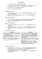
Brain Imaging - Summary
fMRI
● Non-invasive: no health risks associated with procedure, does not use radiation or
involve inserting instruments directly into the brain
○ MRI is super safe (uses non-ionizing radiation unlike CT/PET) but it’s a giant
magnet (B0).
○ Dangers/annoyances
■ No ferromagnetic objects in MRI - EVER → safety risks!
● Bringing anything ferromagnetic into the scanner environment will
cause it to become a projectile, which has been known to kill
people.
■ Radio-Frequency pulses are like a microwave: they can heat up tissue.
■ Loud sounds, Dizziness, Claustrophobia, and Peripheral Nerve
Stimulation
● Provides appealing balance between
○ Temporal resolution (limited): how quickly changes in neural activity within the
brain can be detected
■ Limited by hemodynamic response time / BOLD response
● peak at ~5-6 seconds
○ Spatial resolution (high): ability to identify location of brain activity
○ We could gain as well as lose information by improving resolution!
● The signals we measure depend on oxygen level in the blood
○ They are BOLD (Blood Oxygenation Level Dependent)
■ Provides indirect view of neural activity
■ BOLD measures ratio of oxygenated to deoxygenation hemoglobin in the
blood (in vivo; within a living organism)
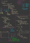
Hemodynamic response: physiological changes that occur in response to neural activity
● This response is slow (~20 seconds) because it depends on complex cascade of
biological metabolic responses such as Cerebral Blood Flow (CBF) and oxygenation
○ causes a stereotypical change in T2* over time
● Underlying process that causes (increased) BOLD response in fMRI
● Typical shape:
MRI scanner: uses strong magnetic fields and radio waves to produce detailed images
● Different field strengths (in Tesla)
○ Higher field strengths generally have a better signal-to-noise ratio, improved
spatial resolution and better contrast between tissues.
■ It has become possible to image cortical processing columns using
smaller and smaller voxels
■ Along with improved sequences, we can scan smaller and faster!
1
,Brain Imaging - Summary
■ Drawbacks of high strengths include cost, complexity and potential safety
concerns
■ What field strength is used may depend on what is examined
● A MRI scan is used to identify abnormalities in the body including the brain (e.g.,
tumors), whereas an fMRI scan is an optimized form of MRI that measures changes in
blood flow/oxygenation in the brain in response to neural activity
MRI Data
● In 3D, values at {x, y, z} → element are voxels: unit that contains
thousands of neurons
● Slicing orientations
○ Sagittal: sideview of head → vertical from nose
○ Coronal: backview → horizontal from top of head
○ Axial: above/top view of head → horizontal from nose
Voxel size and image quality
● Spatial resolution of fMRI depends on voxel size
○ Smaller size provides higher resolution images but require
longer scan times and potentially lower signal-to-noise ratio
○ While it is possible to acquire images of the entire brain at once in fMRI, slicing
provides better image quality and faster acquisition times, making it more
practical
fMRI data
● Contains noise caused by pulsation, movement, veins ‘ringing’
● Various preprocessing steps can be taken to reduce noise
○ Interpolation: correct for motion-related artifacts or align data from different
scans/subjects
fMRI data quality → measured through temporal signal-to-noise ratio (tSNR)
µ (𝑚𝑒𝑎𝑛 𝑖𝑚𝑎𝑔𝑒)
● Formula: tSNR = σ (𝑠𝑡𝑑 𝑜𝑣𝑒𝑟 𝑡𝑖𝑚𝑒)
, calculated at each voxel in an image
○ Reflects strength of BOLD signal relative to noise level in the fMRI data over time
→ not expressed by any SI unit
○ High tSNR: strong signal relative to noise
○ Low tSNR: weak signal relative to noise
● Normal values:
○ ~ 0, air, (μ close to 0)
○ > 40, noisy gray matter (more noise, higher σ)
○ > 80, high-signal gray matter (less noise, lower σ)
○ > 100, white matter (almost no noise, very low σ)
fMRI images as data
𝐼 (𝑖𝑚𝑎𝑔𝑒 𝑎𝑡 𝑡𝑖𝑚𝑒𝑝𝑜𝑖𝑛𝑡 𝑖)
● Percentage signal change (%sc) = 100 * 𝑖
µ (𝑚𝑒𝑎𝑛 𝑖𝑚𝑎𝑔𝑒)
2
,Brain Imaging - Summary
● Normal values
○ 0.2%-1% (normal fMRI experiment @ 3T),
○ 1%-3% (finger-tapping in primary motor cortex @ 3T)
○ 1%-7% (finger-tapping in primary motor cortex @ 7T)
○ 7%-20% (voxel in a large vein in primary cortex @ 7T, where μ is very low)
● Breathing is an example of a factor that can cause signal changes in fMRI images
● Each image represents the BOLD signal in the brain at a particular point in time
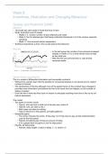
Brain imaging: two major categories
● Structural (T1) images → measures of anatomy and diagnosis of injury → brain structure
○ High spatial resolution
○ Low temporary resolution
■ No temporal resolution actually because they're just a static image
○ Since they have such high spatial resolution they can be used to distinguish
between different tissue types (e.g., separate between grey and white matter)
○ MRI, PET, CAT
● Functional (T2*) images → measures metabolic activity over time → brain function
○ Lower spatial resolution (they're much blurrier than their structural counterparts)
○ Higher temporal resolution and they can therefore be used to relate changes in
signal to an experimental manipulation
○ fMRI, PET, EEG, MEG
Brain imaging modalities
● X-axis: temporal resolution
○ High to low: left to right
● Y-axis: spatial resolution
○ High to low: BOLD fmri, remaining techniques
are similar
Main goals fMRI analysis
● Localization: which regions of the brain are active during a specific task?
● Connectivity: determine how different brain regions are functionally related to one
another → correlations across measured values across time
● Prediction: use a person’s brain activity to predict their perception, behavior or health
status
Blob: cluster of activated brain regions that show increased BOLD signal in response to a
particular task/stimulus
● Size and location give information about the specific brain regions involved in this
task/stimulus
3
, Brain Imaging - Summary
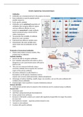
Brain components: small to large
● Neurons → axion carries signal from cellbody to synapses and then to dendrites of other
neurons
● Glia cells → provide protection and support for neurons via schwann cells
○ Majority of the cells in the brain
● Vasculature: network of blood vessels supplying brain with oxygen and glucose
○ From heart to body, back to the heart:
■ Arteries, arterioles, capillaries, venules, veins
● Gray matter: outer layer of the brain (cerebral cortex) → processes and integrates
information
● White matter: inner layer of the brain → allows information to be transmitted across
different parts of the brain
○ Contains all the axon bundles, and these contain a major portion of the fat in the
brain
Broadest brain part separation
● 4 lobes that form cerebral cortex:
○ Frontal lobe → Movement
■ Progressively more abstract,
Higher order control when
moving from posterior (caudal)
to the anterior (rostral) part
○ Parietal lobe → Language, touch
○ Temporal lobe → Learning
○ Occipital lobe → Vision
● Cerebellum → Balance, coordination
● Brain stem → Most important functions
The somatotopic maps of somatosensory and motor cortex are grossly, but not perfectly, aligned
on opposite banks of the central sulcus.
4
fMRI
● Non-invasive: no health risks associated with procedure, does not use radiation or
involve inserting instruments directly into the brain
○ MRI is super safe (uses non-ionizing radiation unlike CT/PET) but it’s a giant
magnet (B0).
○ Dangers/annoyances
■ No ferromagnetic objects in MRI - EVER → safety risks!
● Bringing anything ferromagnetic into the scanner environment will
cause it to become a projectile, which has been known to kill
people.
■ Radio-Frequency pulses are like a microwave: they can heat up tissue.
■ Loud sounds, Dizziness, Claustrophobia, and Peripheral Nerve
Stimulation
● Provides appealing balance between
○ Temporal resolution (limited): how quickly changes in neural activity within the
brain can be detected
■ Limited by hemodynamic response time / BOLD response
● peak at ~5-6 seconds
○ Spatial resolution (high): ability to identify location of brain activity
○ We could gain as well as lose information by improving resolution!
● The signals we measure depend on oxygen level in the blood
○ They are BOLD (Blood Oxygenation Level Dependent)
■ Provides indirect view of neural activity
■ BOLD measures ratio of oxygenated to deoxygenation hemoglobin in the
blood (in vivo; within a living organism)
Hemodynamic response: physiological changes that occur in response to neural activity
● This response is slow (~20 seconds) because it depends on complex cascade of
biological metabolic responses such as Cerebral Blood Flow (CBF) and oxygenation
○ causes a stereotypical change in T2* over time
● Underlying process that causes (increased) BOLD response in fMRI
● Typical shape:
MRI scanner: uses strong magnetic fields and radio waves to produce detailed images
● Different field strengths (in Tesla)
○ Higher field strengths generally have a better signal-to-noise ratio, improved
spatial resolution and better contrast between tissues.
■ It has become possible to image cortical processing columns using
smaller and smaller voxels
■ Along with improved sequences, we can scan smaller and faster!
1
,Brain Imaging - Summary
■ Drawbacks of high strengths include cost, complexity and potential safety
concerns
■ What field strength is used may depend on what is examined
● A MRI scan is used to identify abnormalities in the body including the brain (e.g.,
tumors), whereas an fMRI scan is an optimized form of MRI that measures changes in
blood flow/oxygenation in the brain in response to neural activity
MRI Data
● In 3D, values at {x, y, z} → element are voxels: unit that contains
thousands of neurons
● Slicing orientations
○ Sagittal: sideview of head → vertical from nose
○ Coronal: backview → horizontal from top of head
○ Axial: above/top view of head → horizontal from nose
Voxel size and image quality
● Spatial resolution of fMRI depends on voxel size
○ Smaller size provides higher resolution images but require
longer scan times and potentially lower signal-to-noise ratio
○ While it is possible to acquire images of the entire brain at once in fMRI, slicing
provides better image quality and faster acquisition times, making it more
practical
fMRI data
● Contains noise caused by pulsation, movement, veins ‘ringing’
● Various preprocessing steps can be taken to reduce noise
○ Interpolation: correct for motion-related artifacts or align data from different
scans/subjects
fMRI data quality → measured through temporal signal-to-noise ratio (tSNR)
µ (𝑚𝑒𝑎𝑛 𝑖𝑚𝑎𝑔𝑒)
● Formula: tSNR = σ (𝑠𝑡𝑑 𝑜𝑣𝑒𝑟 𝑡𝑖𝑚𝑒)
, calculated at each voxel in an image
○ Reflects strength of BOLD signal relative to noise level in the fMRI data over time
→ not expressed by any SI unit
○ High tSNR: strong signal relative to noise
○ Low tSNR: weak signal relative to noise
● Normal values:
○ ~ 0, air, (μ close to 0)
○ > 40, noisy gray matter (more noise, higher σ)
○ > 80, high-signal gray matter (less noise, lower σ)
○ > 100, white matter (almost no noise, very low σ)
fMRI images as data
𝐼 (𝑖𝑚𝑎𝑔𝑒 𝑎𝑡 𝑡𝑖𝑚𝑒𝑝𝑜𝑖𝑛𝑡 𝑖)
● Percentage signal change (%sc) = 100 * 𝑖
µ (𝑚𝑒𝑎𝑛 𝑖𝑚𝑎𝑔𝑒)
2
,Brain Imaging - Summary
● Normal values
○ 0.2%-1% (normal fMRI experiment @ 3T),
○ 1%-3% (finger-tapping in primary motor cortex @ 3T)
○ 1%-7% (finger-tapping in primary motor cortex @ 7T)
○ 7%-20% (voxel in a large vein in primary cortex @ 7T, where μ is very low)
● Breathing is an example of a factor that can cause signal changes in fMRI images
● Each image represents the BOLD signal in the brain at a particular point in time
Brain imaging: two major categories
● Structural (T1) images → measures of anatomy and diagnosis of injury → brain structure
○ High spatial resolution
○ Low temporary resolution
■ No temporal resolution actually because they're just a static image
○ Since they have such high spatial resolution they can be used to distinguish
between different tissue types (e.g., separate between grey and white matter)
○ MRI, PET, CAT
● Functional (T2*) images → measures metabolic activity over time → brain function
○ Lower spatial resolution (they're much blurrier than their structural counterparts)
○ Higher temporal resolution and they can therefore be used to relate changes in
signal to an experimental manipulation
○ fMRI, PET, EEG, MEG
Brain imaging modalities
● X-axis: temporal resolution
○ High to low: left to right
● Y-axis: spatial resolution
○ High to low: BOLD fmri, remaining techniques
are similar
Main goals fMRI analysis
● Localization: which regions of the brain are active during a specific task?
● Connectivity: determine how different brain regions are functionally related to one
another → correlations across measured values across time
● Prediction: use a person’s brain activity to predict their perception, behavior or health
status
Blob: cluster of activated brain regions that show increased BOLD signal in response to a
particular task/stimulus
● Size and location give information about the specific brain regions involved in this
task/stimulus
3
, Brain Imaging - Summary
Brain components: small to large
● Neurons → axion carries signal from cellbody to synapses and then to dendrites of other
neurons
● Glia cells → provide protection and support for neurons via schwann cells
○ Majority of the cells in the brain
● Vasculature: network of blood vessels supplying brain with oxygen and glucose
○ From heart to body, back to the heart:
■ Arteries, arterioles, capillaries, venules, veins
● Gray matter: outer layer of the brain (cerebral cortex) → processes and integrates
information
● White matter: inner layer of the brain → allows information to be transmitted across
different parts of the brain
○ Contains all the axon bundles, and these contain a major portion of the fat in the
brain
Broadest brain part separation
● 4 lobes that form cerebral cortex:
○ Frontal lobe → Movement
■ Progressively more abstract,
Higher order control when
moving from posterior (caudal)
to the anterior (rostral) part
○ Parietal lobe → Language, touch
○ Temporal lobe → Learning
○ Occipital lobe → Vision
● Cerebellum → Balance, coordination
● Brain stem → Most important functions
The somatotopic maps of somatosensory and motor cortex are grossly, but not perfectly, aligned
on opposite banks of the central sulcus.
4