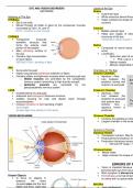
EYE AND VISION DISORDERS Layers of the Eye
MIDTERMS Sclera
Outermost layer
Anatomy of The Eye White avascular dense fibrou
EYEBALL Helps maintain the shape of t
Sits in the orbit contents
Moved through all fields of gaze by the extraocular muscles
innervated by CN II, IV, and VI Choroid
CV III = involves in size of pupil Middle vascular layer
Helps give supply of blood
CORNEA structures of the eye
Transparent avascular *attached to ciliary muscle = l
domelike structure that
forms the anterior most Retina
portion of the eyeball Composed of neural tissue
Main refracting surface of nerve
the eye Landmarks:
Contains high concentration o Optic disc: point of en
of nerve fibers Pink, oval or c
REFRACTION = bending of light o Retinal vessels
o Macula: responsible fo
IRIS
Surrounds the pupil Chambers of The Eye
Highly vascularized pigmented collection of fibers Anterior Chamber:
Contains dilator and sphincter muscles which controls pupil size Aqueous – filled
o Dilator muscles are controlled by the sympathetic Lies between the
nervous system (everything is high and dry except) posterior cornea and
o Sphincter muscles are controlled by the anterior iris and pupil
parasympathetic nervous system
LENS Posterior Chamber
located behind iris and pupil Aqueous – filled
avascular and transparent biconvex structure Lies between the
enables focusing for near and distant vision through posterior iris and pupil
accommodation and anterior lens
involves refraction of light (bending of light) Anterior lens =
focal length aqueous humor is
focuses light rays directly on the retina produced
VISION MECHANISM Vitreous Chamber
Contains the gelatinous vitreo
Largest chamber in the ocula
Humors of The Eye
Aqueous Humor
Transparent nutrient- filled flu
Produced by the posterior ch
Production influences intraoc
o Normal IOP range = 1
Vitreous Humor
Mostly water
Helps maintain the shape of
ERRORS OF R
Vision is impaired because
prevents light rays from focus
Main symptom is blurred vis
Distant Objects Emmetropia: a normal refracti
to focus on objects in on retina with no optical defec
distance, the ciliary
muscles relax and the lens MYOPIA
flatten and thins. Light rays
, ASTIGMATISM
Cause: irregular corneal curve Medical Management:
Irregularity causes the GOAL: maintain a safe level of IOP
incoming light to be bent Pilocarpine; Carbachol
unequally o Cholinergic agents (m
o MOA: opens the trabe
Clinical Manifestations: pupils allowing increas
Headaches o S/E: difficulty seeing in
Distorted vision/ Blurred vision *Miotics = pupil constr
Eye strain Acetazolamide (Diamox)
Squinting o Carbonic Anhydrase I
Difficulty driving at night o MOA: decreases aque
o S/E: GI upset, impoten
Management: Timolol
Prescription eyeglasses o Beta – blocker
LASIK (Laser – assisted in situ keratomileusis) o MOA: decreases aque
o S/E: bradycardia, hyp
LASIK **If not increased level of IOP is not
Surgical procedure optic nerve, thereby causing blindnes
Low to moderate amounts of
myopia or hyperopia, with or Laser Management
without astigmatism Laser trabeculoplasty:
- A laser beam is applied to t
Photorefractive keratectomy (PRK) surface of the trabecular mes
Indications open the intratrabecular spa
o Low to moderate amounts widen the canal of S
of myopia or hyperopia, promoting outflow of aqueou
with or without and decreasing IOP.
astigmatism
o Insufficient corneal Indications:
thickness - Open-angle glaucoma
adequately controlled on
GLAUCOMA tolerated medical therapy
A group of ocular condition characterized by elevated IOP - Open-angle glaucoma in
Common among people older than 40 years’ old which compliance with
No cure but treatable therapy is less than optim
practical, social, or e
reasons or if the medical
effect profile is co
unfavorable for the patien
- Open-angle glaucoma i
medical treatment as initia
Surgical Treatment
Trabeculectomy:
Surgical creation of an openin
fistula in the trabecular mesh
to drain aqueous humor from
anterior chamber to
subconjunctival space
*Progressive eye disorder GOAL: create the right amou
*IOP: influenced by the production of aqueous humor flow without causing overfiltra
Aqueous Humor is produced in the Indications:
ciliary body. From the posterior POAG
chamber, the aqueous humor PCAG unresponsive to iridoto
enters the anterior chamber through Childhood glaucomas
the pupil and drains through the
trabecular meshwork into the canal
of Schlemm, normal flow depends
on the intact drainage system and an Primary Closed Angle Glaucom
open angle (45 - degree). reduction in the outflow
aqueous humor resulting
angle closure