Het vrije onderste lidmaat III
Eerste element: de beenderen:
LET OP!: bij voet lengte-as = horizontaal
dorsoplantaire-as = verticaal
laterolaterale as => blijft behouden
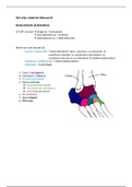
Skelet van voet bestaat uit:
- tarsus (=voetwortel): 7 korte beenderen: talus, calcaneus, os naviculare, os
cuneiforme mediale, os cuneiforme intermedium, os
cuneiforme laterale en os cuboideum (5 middenvoetbeentjes)
- metatarsus (middenvoet): 5 kleine pijpbeenderen
- phalangen: 14 phalangen
, Talus (= sprongbeen):
- Tibia en fibula rusten op talus
- Vormt met tibia en fibula een relatief beweeglijk gewricht
- kop = nr distaal gericht
- plantair zijn er 3 articulaire vlakjes
- lateraal is er proc. lateralis tali
Calcaneus (= hielbeen):
- Facies articularis talaris anterior, media, posterior
- Tuber calcanei vormt beenderige ondergrond van hiel
- mediale kant: sustentaculum tali met de sulcus tendinis musculi flexoris
hallucis longus
- laterale kant: proc. trochlearis met de sulcus tendinis musculi peronei longi
- onderaan: proc. laterale en mediale van het tuber calcanei
- voorkant: facies articularis cuboidea
Os naviculare (pedis):
- Proximaal: facies articularis talaris (concaaf)
- Distaal: articulaire zijde met 3 deeloppn articuleren elk met een os
cuneiforme
- Mediaal: tuberositas ossis navicularis
- Lateraal: kleine opp voor het os cuboideum
Ossa cuneïforme:
3 wiggebeentjes:
os cuneiforme mediale
os cuneiforme intermedium
os cuneiforme laterale
Os cuboideum:
- Laterale zijde: sulcus tendinis musculi peronei longi
- Onderzijde: tuberositas ossis cuboidei
- Mediale zijde: gewrichtsvlakken voor os cuneiforme laterale en os naviculare
- Achterzijde: zadelvormige vlak voor calcaneus
- Voorzijde: een in 2 verdeeld gewrichtsvlak vr de metatarsaalbeentjes IV en V
Ossa metatarsi:
- Algemene vorm: - proximale basis articuleert met beenderen van voetwortel
- een corpus
- distale caput
- Verschil met metacarpalia: - zijn slanker
- dragen duidelijke driehoekige facies articularis
op hun basis
Metatarsale I: - Laterale en plataire zijde: tuberositas ossis metatarsalis I
- Articuleert met os cunïforme mediale
Metatarsale II: - Mediale zijde: articulaire oppervlakte voor os cuneiforme
mediale
- Proximaal: articulair opp met os cuneïforme intermedium
- basis articuleert met basis van metatarsaal III
Metatarsale III: - Articuleert aan proximale zijde met os cuneïforme laterale
- Articuleert mediaal met basis van metatarsaal III
- Articuleert lateraal met basis van metatarsaal V
Eerste element: de beenderen:
LET OP!: bij voet lengte-as = horizontaal
dorsoplantaire-as = verticaal
laterolaterale as => blijft behouden
Skelet van voet bestaat uit:
- tarsus (=voetwortel): 7 korte beenderen: talus, calcaneus, os naviculare, os
cuneiforme mediale, os cuneiforme intermedium, os
cuneiforme laterale en os cuboideum (5 middenvoetbeentjes)
- metatarsus (middenvoet): 5 kleine pijpbeenderen
- phalangen: 14 phalangen
, Talus (= sprongbeen):
- Tibia en fibula rusten op talus
- Vormt met tibia en fibula een relatief beweeglijk gewricht
- kop = nr distaal gericht
- plantair zijn er 3 articulaire vlakjes
- lateraal is er proc. lateralis tali
Calcaneus (= hielbeen):
- Facies articularis talaris anterior, media, posterior
- Tuber calcanei vormt beenderige ondergrond van hiel
- mediale kant: sustentaculum tali met de sulcus tendinis musculi flexoris
hallucis longus
- laterale kant: proc. trochlearis met de sulcus tendinis musculi peronei longi
- onderaan: proc. laterale en mediale van het tuber calcanei
- voorkant: facies articularis cuboidea
Os naviculare (pedis):
- Proximaal: facies articularis talaris (concaaf)
- Distaal: articulaire zijde met 3 deeloppn articuleren elk met een os
cuneiforme
- Mediaal: tuberositas ossis navicularis
- Lateraal: kleine opp voor het os cuboideum
Ossa cuneïforme:
3 wiggebeentjes:
os cuneiforme mediale
os cuneiforme intermedium
os cuneiforme laterale
Os cuboideum:
- Laterale zijde: sulcus tendinis musculi peronei longi
- Onderzijde: tuberositas ossis cuboidei
- Mediale zijde: gewrichtsvlakken voor os cuneiforme laterale en os naviculare
- Achterzijde: zadelvormige vlak voor calcaneus
- Voorzijde: een in 2 verdeeld gewrichtsvlak vr de metatarsaalbeentjes IV en V
Ossa metatarsi:
- Algemene vorm: - proximale basis articuleert met beenderen van voetwortel
- een corpus
- distale caput
- Verschil met metacarpalia: - zijn slanker
- dragen duidelijke driehoekige facies articularis
op hun basis
Metatarsale I: - Laterale en plataire zijde: tuberositas ossis metatarsalis I
- Articuleert met os cunïforme mediale
Metatarsale II: - Mediale zijde: articulaire oppervlakte voor os cuneiforme
mediale
- Proximaal: articulair opp met os cuneïforme intermedium
- basis articuleert met basis van metatarsaal III
Metatarsale III: - Articuleert aan proximale zijde met os cuneïforme laterale
- Articuleert mediaal met basis van metatarsaal III
- Articuleert lateraal met basis van metatarsaal V