PECTORAL REGION
, INTRODUCTION
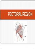
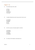
• Pectoral region lies in front of trunk anterior to the thoracic
cage (pector = breast or pectus = chest in Latin).
• It connects the upper limb and anterolateral part of thoracic
wall.
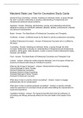
• Major structure of pectoral region:
– Mammary gland
– Muscles
Pectoralis major
Subclavius
Pectoralis minor
Serratus anterior
– Pectoral and clavipectoral fascia
2
, INTRODUCTION
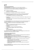
• Gross anatomy of the pectoral
region should be studied in the
following sequence:
Surface landmarks →
Superficial fascia →
Deep fascia →
Muscles (origin, insertion, nerve
supply, action, clinical testing)
3
, SURFACE LANDMARKS
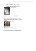
• Surface landmarks are useful for clinical
integration of the gross anatomy.
• Only few of the structures present deep to the
skin can be palpated (felt) or seen as surface
projection. With the help of these structures, all
other structures can be approximately located
and examined.
• These surface landmarks also help for
surgeries.
4
, INTRODUCTION
• Pectoral region lies in front of trunk anterior to the thoracic
cage (pector = breast or pectus = chest in Latin).
• It connects the upper limb and anterolateral part of thoracic
wall.
• Major structure of pectoral region:
– Mammary gland
– Muscles
Pectoralis major
Subclavius
Pectoralis minor
Serratus anterior
– Pectoral and clavipectoral fascia
2
, INTRODUCTION
• Gross anatomy of the pectoral
region should be studied in the
following sequence:
Surface landmarks →
Superficial fascia →
Deep fascia →
Muscles (origin, insertion, nerve
supply, action, clinical testing)
3
, SURFACE LANDMARKS
• Surface landmarks are useful for clinical
integration of the gross anatomy.
• Only few of the structures present deep to the
skin can be palpated (felt) or seen as surface
projection. With the help of these structures, all
other structures can be approximately located
and examined.
• These surface landmarks also help for
surgeries.
4