

CCRN Exam Cram review
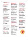
CARDIOVASCULAR REVIEW STUDY GUIDE CCRN Exam Cram Heart Sounds S1, S3, S4: Heard at apex S2: Heard at base S3: r/t heart failure S4: r/t MI, HTN, ventricular hypertrophy, & aortic stenosis ……………………………… Mitral stenosis is associated with atrial fibrillation due to atrial enlargement. ……………………………… Stenosis occurs with an OPEN valve. Insufficiency occurs with a CLOSED valve. ……………………………… NSTEMI: + troponin, ST depression, unrelenting chest pain. STEMI: + troponin, ST elevation, unrelenting chest pain. Prinzmetal’s (Variant) Angina: - troponin, nitroglycerin relieves chest pain and returns ST segment to normal Management of Acute Chest Pain STAT EKG done and read within 10 minutes, 325mg chewable aspirin ASAP, anticoagulant therapy, antiplatelet therapy, beta blocker administration. Use metoprolol, NOT propranolol. Treat chest pain with morphine or nitroglycerin. If symptoms have been present 12 hours, prepare for PCI. Door to balloon in 90 minutes. Door to fibrinolytic therapy in 30 minutes. ……………………………… After PCI Evidence of successful reperfusion includes chest pain relief, normalized ST segment, and reperfusion arrhythmias (VT, VF). Troponin and CK-MB remain elevated after reperfusion! Complications of PCI Monitor for signs of re-occlusion… chest pain and ST elevation similar to before PCI. Monitor for vasovagal response during sheath removal. Administer atropine and fluids, hold nitrates. Monitor for signs of retroperitoneal bleeding… back pain and hypotension. HTN Emergency BP 180/110 with evidence of end-organ damage. Needs critical care admission and emergent lowering of BP. Biggest risk is STROKE. Nitroprusside Drip: preload and afterload reducer. Assess for cyanide toxicity (thiocyanate)... mental status changes, tachycardia, seizure, need for increase in dose, metabolic acidosis. Labetalol IV Push: Intermittent IV pushes preferred over drip due to the possibility of continuing drip past max of 300mg. ……………………………… Acute Peripheral Vascular Insufficiency Normal ankle-brachial index is 1. Put bed in reverse trendelenburg to increase blood flow. Do NOT elevate the extremity! Medical Management: tPA, thrombolytics, heparin, anticoagulants, antiplatelets, vasodilators. 1 QT Prolongation Normal QT interval is 0.36 to 0.4 seconds. Prolongation of the QT interval can lead to Torsades de Pointes! Treatment of Torsades de Pointes is magnesium. Drugs that prolong QT: amiodarone, quinidine, haloperidol, procainamide. Electrolytes that prolong QT: hypokalemia, hypocalcemia, hypomagnesemia. ……………………………… 3 Pacemaker Malfunctions Failure to Pace: No pacer spike when expected. Failure to Capture: Pacer spike occurs without QRS complex. Failure to Sense: Pacing in native beats. ……………………………… Heart Failure Elevated LVEDP. High intracardiac pressures & decreased CO. Systolic Heart Failure: Ejection problem, EF 40%. Dilated ventricle. S3 heart sound. Large, dilated heart or normal heart size on CT scan. Treatment: Beta blockers, ACE/ARBs, positive inotropes. Contraindicated: Calcium channel blockers & negative inotropes. VS Diastolic Heart Failure: Filling problem, EF 50%. Thick, hypertrophic ventricle. S4 heart sound with hypertension. Normal heart size on CT scan. Treatment: Beta blockers, ACE/ARBs, calcium channel blockers. Contraindicated: Positive inotropes (no digoxin, dopamine, or dobutamine for these patients). ……………………………… Systolic heart failure can cause valvular insufficiency r/t a dilated ventricle. Both systolic and diastolic heart failure cause pulmonary edema r/t poor ventricular emptying. Symptoms of Right-Sided Heart Failure: Hepatomegaly, splenomegaly, dependent edema, increased CVP, tricuspid insufficiency, abdominal pain. VS Symptoms of Left-Sided Heart Failure: Orthopnea, dyspnea, tachypnea, hypoxemia, tachycardia, crackles, cough with pink/frothy sputum, diaphoresis, anxiety, confusion, increased PAP & PAOP. ……………………………… NYHA Heart Failure Classification Class I: Symptoms caused by extraordinary activity. Class II: Symptoms caused by ordinary activity. Class III: Symptoms caused by minimal activity (such as walking to the bathroom). Class IV: Symptoms at rest. Cardiogenic Shock Extreme decrease in stroke volume r/t systolic dysfunction. Increased PAOP r/t increased LV preload. Increased SVR r/t compensatory vasoconstriction. Decreased CO means that perfusion to organs is inadequate. Narrow pulse pressure. Treatment: Give positive inotropes! Give norepinephrine, dopamine 10 mcg, dobutamine, or milrinone. Also, reduce preload & afterload with vasodilators in conjunction with positive inotropes, IABP, or VAD. Ventricular Assist Device (VAD) Used in the management of left ventricular heart failure, cardiogenic shock, and cardiomyopathy. Used for patients awaiting a heart transplant. Intra Aortic Balloon Pump (IABP) Inflates during diastole to increase myocardial oxygen supply. Deflates during systole to reduce left ventricular afterload. Post-Op CABG Nursing Care Monitor for complications of cardiac tamponade and pericarditis. Do not clamp or milk chest tubes, maintain dependent drainage with NO LOOPS IN TUBING. Monitor for chest tube output 100mL for two consecutive hours. ……………………………… Valve Replacement High risk for blood clots, need anticoagulation! Avoid a drop in preload! May result in dangerous hypotension. Conduction disturbances r/t surgery near SA/AV nodes. May need a temporary or permanent pacemaker. ……………………………… Cardiac Tamponade Symptoms: Hypotension, JVD, muffled heart tones, equalization of CVP/PAOP, narrowed pulse pressure, pulsus paradoxus, enlarged cardiac silhouette. Pulsus Paradoxus is a drop in SBP during inspiration. Cardiac Trauma The aortic valve is most likely to be ruptured because it is most anterior. Pain is worse with inspiration. ST elevation present in the area of injury only. Worse outcome than pericarditis. VS Pericarditis ST elevation in ALL LEADS (aka “global” ST elevation). Pain is worse with inspiration. Dressler’s Syndrome is pericarditis that occurs after MI, surgery, or injury. ……………………………… Thoracic Aneurysms Aneurysms 6 cm needs surgery and blood pressure control with a labetalol drip. Sudden tearing, ripping pain in chest, radiating to shoulders/back, cough, and widening mediastinum on X-ray indicate a RUPTURE, which is a surgical emergency! PULMONARY REVIEW CCRN Exam Cram → What is the clinical indicator of ventilation? PCO2! → What is the primary muscle of ventilation? The diaphragm! → Normal ventilation is about 4L/min. → Normal SVO2 is 60-75% → Normal PO2 is 80-100 mmHg, 80 is hypoxemia. → The goal for most critically ill patients is O2 sat 90%, PO2 60 mmHG. ……………………………… Position the patient’s GOOD LUNG DOWN to prevent hypoxemia. You want to INCREASE PERFUSION to the GOOD LUNG by positioning it DOWN. ……………………………… Static Compliance: Elasticity of the lung. Dynamic Compliance: Elasticity of the airways. In asthma, static compliance is ok, dynamic compliance is decreased. Noninvasive Ventilation CPAP: For patients with hypoxemic respiratory failure. BiPAP: For patients with hypoxemic and/or hypercapnic respiratory failure. Contraindications to NIV: Hemodynamic instability or life-threatening arrhythmias, copious secretions, high risk for aspiration, impaired mental status (unable to protect airway!), suspected pneumothorax, inability to cooperate, life-threatening refractory hypoxemia. A patient with these contraindications may need INTUBATION instead of NIV! ……………………………… Carbon Monoxide Poisoning Do not use pulse oximetry to monitor O2 status, the pulse ox cannot differentiate between CO and O2! Treat with 100% FiO2 until symptoms resolve and CO level is 10%. Status Asthmaticus Severe airway narrowing refractory to bronchodilators. Wheezing → Decreased Breath Sounds → Absent Breath Sounds… THIS IS BAD! Condition is getting worse. Starts with respiratory alkalosis r/t hyperventilation, when the patient tires out and respiratory rate drops respiratory acidosis occurs r/t hypoventilation. THIS IS BAD! Condition is getting worse. Measure presenting peak flow rate. Intubate if… respiratory acidosis, severe hypoxemia, silent chest, decrease in level of consciousness. Ventilator Management for Asthma: Use low respiratory rate to increase exhalation time. Increase inspiration:expiration ratio to 1:3-4 to allow time for optimal exhalation and prevent auto-PEEP. Use low tidal volumes to prevent auto-PEEP. COPD Exhalation is HARD! Causes air trapping and auto-PEEP. V/Q mismatch r/t a problem with ventilation and increased PCO2. Treatment: Increase FiO2 to keep O2 sat 90% or PO2 60. Use bronchodilators, corticosteroids, antibiotics for pneumonia, BiPAP or ventilator. ……………………………… A PULMONARY EMBOLISM results in DEAD SPACE. It responds to O2 therapy, so GIVE O2. VS ARDS is a SHUNT and will NOT respond to increased FIO2, it also needs PEEP! PEEP prevents alveolar collapse and increases alveolar recruitment. A SHUNT (ARDS!) is movement of blood from the right side of the heart to the left side without getting oxygenated. Pulmonary Embolism V/Q mismatch, increased PAP. Pulmonary angiography is the gold standard for diagnosis. Symptoms: dyspnea, tachypnea, tachycardia, cough, crackles, anxiety, respiratory alkalosis. PETECHIAE if the embolism is a FAT EMBOLISM. Treatment: Fluids! Anticoagulation (heparin drip or low-molecular weight heparin), fibrinolytic therapy for all patients with hemodynamic compromise with low risk for bleeding. ……………………………… ARDS Inflammation causes increased permeability of the pulmonary capillary membrane. Proteinaceous fluid leaks into the interstitial and alveolar spaces. Alveolar Type II cells are damaged, which results in decreased surfactant production and MASSIVE ATELECTASIS! This creates a SHUNT. FiO2 is not enough, ARDS needs PEEP! PEEP increases alveolar recruitment to treat refractory hypoxemia. Refractory hypoxemia is hypoxemia even when FiO2 is 100%. Symptoms: Bilateral infiltrates on chest X-ray. PAOP 18 because this is a lung problem, not a heart problem. Decreased lung compliance, increased work of breathing, Decreased functional residual capacity. Pulmonary edema, crackles. Treatment: Intubate and ventilate with PEEP ≥ to 15. Monitor for barotrauma and decreased CO, but DO NOT STOP PEEP! Do not disconnect from the ventilator circuit because alveolar recruitment will be lost and is not easily corrected! Limit tidal volume to 4-6 mL/kg to prevent volutrauma! (Normal tidal volume setting is 8-10 mL/kg). Nutritional support is important! Prone positioning.
Document information
- Uploaded on
- December 2, 2023
- Number of pages
- 30
- Written in
- 2023/2024
- Type
- Exam (elaborations)
- Contains
- Questions & answers


