

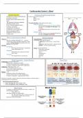
Cardiovascular System 1 - Blood
Learning Outcomes: Overview of Cardiovascular System:
- Components of blood and blood types Cardiovascular system consists of:
- Location of the heart - Heart
- Internal and external features of the - Blood vessels
- Blood
heart
- The function of the heart chambers Two major divisions of the cardiovascular system:
- Function of heart valves - Pulmonary Circuit.
- Flow of blood through the heart o right side of heart receives deoxygenated blood
- Coronary circulation from the body and pumps it to the lungs for gas
- Components of cardiocyte exchange
- Conduction system of the heart o oxygenated blood returns to the left side of the
- Electrocardiogram (ECG) heart
- Systemic Circuit:
- Regulation of heart activity
o left side of heart receives oxygenated blood from
- Cardiac cycle the lungs and pumps it to the body
o deoxygenated blood returns to the right side of the
What is it and functions of Blood? heart Blood Components
What is it? Formed Elements (living blood cells) 45%
- Blood is an alkaline liquid (pH 7.35 – 7.45) that All originate in the red bone marrow
contains both cellular and liquid components, which - erythrocytes (red blood cells) – carry O2 and
consist of formed elements (living blood cells) and CO2
plasma (non-living fluid) - leukocytes (white blood cells) – immune function
Functions: - platelets – involved in blood clotting
- Distribution
o Delivering O2 and nutrients, transporting Plasma: non-living fluid 55%
wastes and hormones, etc. - H2O (main constituent)
- Regulation - dissolved proteins
o Maintaining body temperature, pH and - glucose
fluid volume - clotting factors
- Protection - electrolytes
o Preventing blood loss and infection - hormones
- O2
- CO2
Blood Components – Formed Elements Blood Types
Erythrocytes: (Red blood cells) 44%
- Flattened discs with depressed centre
- Contains haemoglobin (a gas transporting protein)
- No nuclei or organelles (obtain ATP via glycolysis)
Leukocytes: (White blood cells) -<1%
- Lots of different types/size range
- Can leave the cardiovascular system
- Respond to foreign bodies
Platelets: - <1%
- Fragments of larger cells megakaryocytes
- Contain chemicals that act in the clotting process
- No nuclei or organelles – degenerate after 10 days
Erythrocytes normally constitute about 44% of the total volume of a blood sample, a
percentage known as a haematocrit.
Blood Typing
- In addition, the ABO blood group system there is another cell
protein that can be present
o Rhesus (Rh) factor
- Individuals with Rh are termed positive
o E.g. AB+ type blood = have A surface proteins, B
surface proteins and Rh surface proteins
- Important for blood transfusions
, Cardiovascular System 1 – The Heart
The Heart
Located in the medial compartment of the thorax called the
mediastinum.
- Posterior to the sternum
- Anterior to the vertebral column
- Superior to the diaphragm
- Medial to the lungs
Pericardium
Pericardium: double walled sac that encloses the heart
- Superficial part: Fibrous pericardium
o Tough, dense connective tissue which protects, anchors, and prevents
overfilling
- Deep Part: Serous pericardium
Wall Layers o Thin, slippery, two-layer serous membrane which allows smooth gliding
Three layers of the heart wall: ▪ Parietal layer: lines the internal surface of the fibrous pericardium
1. Epicardium: superficial layer ▪ Visceral Layer: covers the external heat surface
2. Myocardium: middle layer = cardiac muscle layer,
makes up bulk of the heart Pericardial Cavity:
3. Endocardium: deep layer, lines the heart chambers and - Contains a film of serous fluid which allows the heart to work in a friction free
valves environment
The Heart Chambers
Four Chambers:
- Two Upper Atria:
o Separated by the thin interatrial septum
▪ Contains the fossa ovalis = foetal remnant
- Two lower Ventricles:
o Separated by thick interventricular septum
o Contain papillary muscles
- Coronary sulcus:
o Encircles the junction between atria and ventricles
o Contains coronary vessels
- Interventricular sulcus:
o Overlies the interventricular septum
o Contains coronary vessels
Fibrous Skeleton
Fibrous skeleton of the heart:
- Surround and support all valves,
prevents from stretching
- Separate atria and ventricles
- Provide attachment for muscle fibres
- Form the basis of electrical
discontinuity between atria and
ventricles
, Cardiovascular System 1 – The Heart
Valves Blood Flow
Atrioventricular valves:
- Operate at one-way valves to direct blood and prevent backflow
- Located between right atrium and ventricle, left atrium and ventricle
- Open/close passively
Semilunar Valves:
- Contains 3 cusps
- Operate at one-way valves to direct blood and prevent backflow
- Located between right ventricle and pulmonary trunk and left ventricle and aorta
- Open/close passively
Pulmonary Circuit:
- Short loop
- Low pressure
- 100% of blood
Systemic Circuit:
Coronary Circulation - Long loop
Arteries: - High pressure
- The heart supplies itself with blood via coronary circulation - 100% of blood
- Left coronary artery (LCA) and right coronary artery (RCA) branch off the
aorta just above the aortic valve
Cardiocytes
Cardiocytes: contractile cells of the heart (muscle tissues)
- Any myocyte of the heart (myocyte is the cells that make up
muscle tissue)
- Contain 1 or 2 nuclei surrounded by glycogen
- Myocardial Infarction (heart attack): cardiac muscle death resulting from - Contain many mitochondria
prolonged coronary blockage - Interconnected by intercalated discs
o Contain desmosomes
Veins: ▪ Mechanical junctions between the cells that
- Great cardiac vein (ant), middle cardiac vein (post.) and small cardiac vein prevent cardiocytes from pulling apart
(ant and post) drain into the coronary sinus → right atrium o Contains gap junctions
▪ Allows ions to flow between cells to spread
action potentials
▪ Allows the myocardium to act as a single
coordinated unit, functional syncytium
Metabolism of Cardiac Muscle
- Cardiocytes are fatigue resistant, do not fatigue for a lifetime
- Cardiac muscle depends almost exclusively on aerobic respiration to make ATP
needed for contraction
o Aerobic respiration = the process of producing cellular energy
involving energy (i.e oxygen + fuel = energy)
- Rich in myoglobin (stored oxygen) and glycogen (stored glucose), while
mitochondria makes up 30% of cell volume
- Adaptable to all organic fuels but generally use fatty acids, glucose, lactic acid
and amino acid
- Due to reliance on aerobic restoration, cardiac muscle is very vulnerable to
oxygen deficiency which can cause myocardial infarction
Learning Outcomes: Overview of Cardiovascular System:
- Components of blood and blood types Cardiovascular system consists of:
- Location of the heart - Heart
- Internal and external features of the - Blood vessels
- Blood
heart
- The function of the heart chambers Two major divisions of the cardiovascular system:
- Function of heart valves - Pulmonary Circuit.
- Flow of blood through the heart o right side of heart receives deoxygenated blood
- Coronary circulation from the body and pumps it to the lungs for gas
- Components of cardiocyte exchange
- Conduction system of the heart o oxygenated blood returns to the left side of the
- Electrocardiogram (ECG) heart
- Systemic Circuit:
- Regulation of heart activity
o left side of heart receives oxygenated blood from
- Cardiac cycle the lungs and pumps it to the body
o deoxygenated blood returns to the right side of the
What is it and functions of Blood? heart Blood Components
What is it? Formed Elements (living blood cells) 45%
- Blood is an alkaline liquid (pH 7.35 – 7.45) that All originate in the red bone marrow
contains both cellular and liquid components, which - erythrocytes (red blood cells) – carry O2 and
consist of formed elements (living blood cells) and CO2
plasma (non-living fluid) - leukocytes (white blood cells) – immune function
Functions: - platelets – involved in blood clotting
- Distribution
o Delivering O2 and nutrients, transporting Plasma: non-living fluid 55%
wastes and hormones, etc. - H2O (main constituent)
- Regulation - dissolved proteins
o Maintaining body temperature, pH and - glucose
fluid volume - clotting factors
- Protection - electrolytes
o Preventing blood loss and infection - hormones
- O2
- CO2
Blood Components – Formed Elements Blood Types
Erythrocytes: (Red blood cells) 44%
- Flattened discs with depressed centre
- Contains haemoglobin (a gas transporting protein)
- No nuclei or organelles (obtain ATP via glycolysis)
Leukocytes: (White blood cells) -<1%
- Lots of different types/size range
- Can leave the cardiovascular system
- Respond to foreign bodies
Platelets: - <1%
- Fragments of larger cells megakaryocytes
- Contain chemicals that act in the clotting process
- No nuclei or organelles – degenerate after 10 days
Erythrocytes normally constitute about 44% of the total volume of a blood sample, a
percentage known as a haematocrit.
Blood Typing
- In addition, the ABO blood group system there is another cell
protein that can be present
o Rhesus (Rh) factor
- Individuals with Rh are termed positive
o E.g. AB+ type blood = have A surface proteins, B
surface proteins and Rh surface proteins
- Important for blood transfusions
, Cardiovascular System 1 – The Heart
The Heart
Located in the medial compartment of the thorax called the
mediastinum.
- Posterior to the sternum
- Anterior to the vertebral column
- Superior to the diaphragm
- Medial to the lungs
Pericardium
Pericardium: double walled sac that encloses the heart
- Superficial part: Fibrous pericardium
o Tough, dense connective tissue which protects, anchors, and prevents
overfilling
- Deep Part: Serous pericardium
Wall Layers o Thin, slippery, two-layer serous membrane which allows smooth gliding
Three layers of the heart wall: ▪ Parietal layer: lines the internal surface of the fibrous pericardium
1. Epicardium: superficial layer ▪ Visceral Layer: covers the external heat surface
2. Myocardium: middle layer = cardiac muscle layer,
makes up bulk of the heart Pericardial Cavity:
3. Endocardium: deep layer, lines the heart chambers and - Contains a film of serous fluid which allows the heart to work in a friction free
valves environment
The Heart Chambers
Four Chambers:
- Two Upper Atria:
o Separated by the thin interatrial septum
▪ Contains the fossa ovalis = foetal remnant
- Two lower Ventricles:
o Separated by thick interventricular septum
o Contain papillary muscles
- Coronary sulcus:
o Encircles the junction between atria and ventricles
o Contains coronary vessels
- Interventricular sulcus:
o Overlies the interventricular septum
o Contains coronary vessels
Fibrous Skeleton
Fibrous skeleton of the heart:
- Surround and support all valves,
prevents from stretching
- Separate atria and ventricles
- Provide attachment for muscle fibres
- Form the basis of electrical
discontinuity between atria and
ventricles
, Cardiovascular System 1 – The Heart
Valves Blood Flow
Atrioventricular valves:
- Operate at one-way valves to direct blood and prevent backflow
- Located between right atrium and ventricle, left atrium and ventricle
- Open/close passively
Semilunar Valves:
- Contains 3 cusps
- Operate at one-way valves to direct blood and prevent backflow
- Located between right ventricle and pulmonary trunk and left ventricle and aorta
- Open/close passively
Pulmonary Circuit:
- Short loop
- Low pressure
- 100% of blood
Systemic Circuit:
Coronary Circulation - Long loop
Arteries: - High pressure
- The heart supplies itself with blood via coronary circulation - 100% of blood
- Left coronary artery (LCA) and right coronary artery (RCA) branch off the
aorta just above the aortic valve
Cardiocytes
Cardiocytes: contractile cells of the heart (muscle tissues)
- Any myocyte of the heart (myocyte is the cells that make up
muscle tissue)
- Contain 1 or 2 nuclei surrounded by glycogen
- Myocardial Infarction (heart attack): cardiac muscle death resulting from - Contain many mitochondria
prolonged coronary blockage - Interconnected by intercalated discs
o Contain desmosomes
Veins: ▪ Mechanical junctions between the cells that
- Great cardiac vein (ant), middle cardiac vein (post.) and small cardiac vein prevent cardiocytes from pulling apart
(ant and post) drain into the coronary sinus → right atrium o Contains gap junctions
▪ Allows ions to flow between cells to spread
action potentials
▪ Allows the myocardium to act as a single
coordinated unit, functional syncytium
Metabolism of Cardiac Muscle
- Cardiocytes are fatigue resistant, do not fatigue for a lifetime
- Cardiac muscle depends almost exclusively on aerobic respiration to make ATP
needed for contraction
o Aerobic respiration = the process of producing cellular energy
involving energy (i.e oxygen + fuel = energy)
- Rich in myoglobin (stored oxygen) and glycogen (stored glucose), while
mitochondria makes up 30% of cell volume
- Adaptable to all organic fuels but generally use fatty acids, glucose, lactic acid
and amino acid
- Due to reliance on aerobic restoration, cardiac muscle is very vulnerable to
oxygen deficiency which can cause myocardial infarction


