▪ ▪
Lu 10 A&P
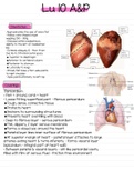
Orientation
• Approximately the size of one’s fist
• Hollow, cone shaped organ
weighing 250 – 350g
• Enclosed within mediastinum,
slightly to the left of mediastinal
line
• Extends obliquely 12 – 14cm from
2nd rib to 5th intercostal space
Superior to diaphragm
Anterior to vertebral column
Posterior to sternum
Laterally to lungs
• Broad, flat base ± 9cm wide points
to right shoulder
• Apex points inferiorly to left hip
Coverings
•Pericardium
• Peri = around, cardi = heart
• Loose fitting superficial part – fibrous pericardium:
Tough, dense, connective tissue
Protects heart
Anchors to surrounding structures
Prevents heart overfilling with blood
• Deep to fibrous layer – serous pericardium:
Thin, slippery, 2 layer serous membrane
Forms a closed sac around the heart
Parietal layer lines inner surface of fibrous pericardium
At superior margin of heart – parietal layer attaches to large
arteries exiting heart & turns inferiorly – forms visceral layer
(epicardium – integral part of heart wall)
• Between parietal & visceral layers – slit-like pericardial cavity
filled with film of serous fluid – friction free environment
, Layers
•3 layers
Epicardium
Myocardium
Endocardium
• Epicardium – visceral layer of
serous
pericardium
• Myocardium – “muscle heart”
Composed of cardiac muscle
Forms bulk of heart
Contracts
Made up of circular bundles of
connective tissue fibers – additional
support for vessels and valves
leaving
the heart
Connective tissue not electrically
excitable – action potentials only
spread
via specific pathways
• Endocardium – “inside the heart”
White sheet of endothelium, rests on thin connective
tissue layer
Lines heart chambers and covers skeleton of valves
Continuous with lining of blood vessels
Lu 10 A&P
Orientation
• Approximately the size of one’s fist
• Hollow, cone shaped organ
weighing 250 – 350g
• Enclosed within mediastinum,
slightly to the left of mediastinal
line
• Extends obliquely 12 – 14cm from
2nd rib to 5th intercostal space
Superior to diaphragm
Anterior to vertebral column
Posterior to sternum
Laterally to lungs
• Broad, flat base ± 9cm wide points
to right shoulder
• Apex points inferiorly to left hip
Coverings
•Pericardium
• Peri = around, cardi = heart
• Loose fitting superficial part – fibrous pericardium:
Tough, dense, connective tissue
Protects heart
Anchors to surrounding structures
Prevents heart overfilling with blood
• Deep to fibrous layer – serous pericardium:
Thin, slippery, 2 layer serous membrane
Forms a closed sac around the heart
Parietal layer lines inner surface of fibrous pericardium
At superior margin of heart – parietal layer attaches to large
arteries exiting heart & turns inferiorly – forms visceral layer
(epicardium – integral part of heart wall)
• Between parietal & visceral layers – slit-like pericardial cavity
filled with film of serous fluid – friction free environment
, Layers
•3 layers
Epicardium
Myocardium
Endocardium
• Epicardium – visceral layer of
serous
pericardium
• Myocardium – “muscle heart”
Composed of cardiac muscle
Forms bulk of heart
Contracts
Made up of circular bundles of
connective tissue fibers – additional
support for vessels and valves
leaving
the heart
Connective tissue not electrically
excitable – action potentials only
spread
via specific pathways
• Endocardium – “inside the heart”
White sheet of endothelium, rests on thin connective
tissue layer
Lines heart chambers and covers skeleton of valves
Continuous with lining of blood vessels