Relias Cardiac dysrhythmia management & pacemakers Study Guide.
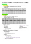
Relias Cardiac dysrhythmia management & pacemakers Study Guide. Cardiac dysrhythmias − Students to review slides 4-29 prior to class: this content will not be covered in NUR 4120 − Normal sinus rhythm • Answers to evaluation of rhythm will always be within normal limits • Rate: 60-100bpm Sinus node dysrhythmias − Sinus bradycardia • HR < 60 bpm • Sinus node creates impulse at slower than normal rate • Characteristics of NSR but a slower rate − Etiology: sinus bradycardia • Sleep • Athletic training • Hypothyroidism • Vagal stimulation ▪ Vomiting, suctioning, pain • Medication ▪ CCB (decrease HR/BP), amiodarone, beta-blockers • Increased intracranial pressure • CAD/Acute MI • Hypoxemia • Altered mental status • Acute decompensated heart failure − Sinus bradycardia: clinical manifestations and management • Clinical manifestations: ▪ SOB altered LOC ▪ Hypotension ▪ EKG changes (ST segment changes PVC’s) • Management: ▪ Resolve causative factors ▪ Atropine 0.5 mg IV every 3-5 minutes Maximum dose of 3 mg Atropine won’t work on a patient with a heart transplant ▪ Emergency transcutaneous pacing ▪ Catecholamines − Sinus tachycardia • HR: 100-120 • Sinus node creates impulse at faster than normal rate • Does not start or stop suddenly − Etiology: sinus tachycardia • Physiologic stress ▪ Acute blood loss, anemia ▪ Shock ▪ Hyper/hypovolemia ▪ Heart failure ▪ Pain ▪ Hypermetabolic states ▪ Fever ▪ Exercise ▪ Anxiety • Medications ▪ Catecholamine ▪ Atropine ▪ Stimulants (caffeine, nicotine) ▪ Illicit drugs (Ecstasy, cocaine) − Sinus tachycardia: clinical manifestations and management • Clinical manifestations ▪ Decreased filling time of heart Reduces cardiac output → Syncope → Hypotension → Acute pulmonary edema (assess lung sounds, diff. breathing) • Management ▪ Abolish the cause ▪ Synchronized cardioversion (hemodynamic instability) ▪ Vagal maneuvers → recharges SA node ▪ Adenosine (only for narrow QRS) ▪ Narrow QRS? Beta-blockers (rare) Calcium-channel blockers (rare) Adenosine ▪ Wide QRS? sotalol, amiodarone ▪ Increased fluid/sodium (POTS) postural orthostatic tachycardia Atrial dysrhythmias − Atrial flutter • Conduction defect in the atrium, filling time is affected, risk = coagulation • Creates atrial rate between 250-400 times/minute (ventricular rate 75-150) • Not all impulses conducted to ventricle: therapeutic block at AV node • 2:1, 3:1, 4:1 • Regular atrial activity • P wave = “saw tooth” appearance • HR > 100 bpm ▪ “uncontrolled” • HR > 150 bpm ▪ “rapid ventricular rate” − Etiology: atrial flutter** • COPD • Pulmonary HTN • Valvular disease • Thyrotoxicosis • Open heart surgery − Atrial flutter: clinical manifestations and management • Clinical manifestations: ▪ Chest pain ▪ Dyspnea ▪ Hypotension • Management: ▪ Electrical cardioversion for unstable patient ▪ See treatment for atrial fibrillation ▪ Medications to slow the ventricular response: Beta blockers Calcium channel blockers Digitalis (digoxin) decreases HR Diltiazem ▪ Usually resolves on own but if it doesn’t resolve within 48 hours, look out for a blood clot/coagulation − Atrial fibrillation (more disorganized than atrial flutter) • Rapid, disorganized and uncoordinated twitching of atrial muscle • Paroxysmal or chronic • Rapid ventricular response; loss of atrial kick (25-30% of cardiac output) • Atrial rate 300-600 BPM • Ventricular rate: 120-200 BPM • Paroxysmal: recurrent; with sudden onset and termination • Persistent: continuous • Permanent: persistent = decision made not to restore or maintain NSR − Etiology: atrial fibrillation • Post-operative period after CABG • Valvular disease • Inflammatory disease (pericarditis) • HTN • CAD, cardiomyopathy • Heart failure • Hyperthyroidism • Pulmonary HTN and embolism • OSA (obstructive sleep apnea) • “holiday heart” (alcohol) • Subarachnoid hemorrhage − Atrial fibrillation: clinical manifestations and diagnosis • Clinical manifestations ▪ Palpitations ▪ Fatigue ▪ SOB ▪ Exercise intolerance ▪ Hemodynamic collapse • Diagnosis ▪ Depends on cause and duration, patient age, symptoms and co-morbidities ▪ 12-lead EKG ▪ Echocardiogram ▪ Thyroid, renal and hepatic function labs ▪ CXR → identify respiratory system problems, lung related ▪ Exercise test ▪ Holter monitoring − Atrial fibrillation: management • Rhythm control vs. rate control • Hemodynamically unstable? ▪ Electrical cardioversion if < 48 hours ▪ > 48 hours: TEE to confirm mural wall thrombus If absent: heparin prior to cardioversion High risk of embolization of atrial thrombi if cardioverted if AF duration > 48 hours Coumadin x 4 weeks after cardioversion Amiodarone, betapace, rhythmol prior to cardioversion ▪ Pharmacologic cardioversion Tikosyn, ibutelid = required patient hospitalization • HR control • Beta blocker ▪ Contraindicated with bronchospasm • Calcium channel blocker.
Written for
- Institution
- Relias Cardiac dysrhythmia management & pacemakers
- Course
- Relias Cardiac dysrhythmia management & pacemakers
Document information
- Uploaded on
- April 5, 2023
- Number of pages
- 17
- Written in
- 2022/2023
- Type
- Exam (elaborations)
- Contains
- Questions & answers
Subjects
- cardiac dysrhyth
-
relias cardiac dysrhythmia management amp pacemakers study guide
-
relias cardiac dysrhythmia management amp pacemakers
-
cardiac dysrhythmia management amp pacemakers study guide
Also available in package deal