

Year 3 Week 1 Physiology
Second lecture
Methods in Skeletal Muscle Physiology Research:
Neuromuscular Function
Measuring muscle strength
in vivo:
● Dynamic strength e.g. 1rm - Doesn’t isolate the muscle (e.g. Leg drive on bench press,
stabiliser muscles)
● Maximum isometric strength - Isolates the muscle (Joints in a fixed position strapped into
a machine
What are the two main determinants of maximal muscle strength?
● Neural activation capacity - Our neuromuscular system becomes more effective and
efficient by learning how to recruit more motor units and muscle fibres to produce more
output resulting in becoming stronger.
● Muscle size
Measuring muscular activation
In vivo:
● Voluntary muscle activation - more invasive method
, Interpolated twitch technique:
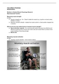
● Isometric rig shown in photo, force transducer used to measure the force of the
quadriceps to try and extend the leg and the knee joint
● Ankle strapped to contraption so cant extend the leg at the knee joint so its an isometric
contraction
● Two large electrodes on thigh supply electrical stimulation from the machine that causes
the muscle fibres in the quadriceps to contract involuntarily
● Then ask him to perform a MVC (maximum voluntary contraction)
● Third red circle is another electrical stimulus after he’s reached a plateau
● This extra force produced is from motor units and muscle fibres that he has not been
able to activate or stimulate him
● Therefore, his voluntary contraction of quads is below 100%
How do we calculate this percentage?
● Compare the superimposed force with the baseline involuntary force
● The smaller the baseline involuntary force, the better
● As he can recruit a higher percentage of his muscle fibres and motor units
● Larger = he cant activate as many motor units in the quadricep
● Young healthy people = about 90%
● Go to the gym for a few months this can increase by approx 5%
Muscle activation: surface EMG (electromyography)
● Less invasive
● Clean the skin, then put electrodes on muscle of interest
● Electrodes record the electrical activity in the muscle
● Electrical signal comes from brain then depolarisation through muscle
● More motor units and muscle fibres that are innovated, the greater electrical signal we
pick up
Second lecture
Methods in Skeletal Muscle Physiology Research:
Neuromuscular Function
Measuring muscle strength
in vivo:
● Dynamic strength e.g. 1rm - Doesn’t isolate the muscle (e.g. Leg drive on bench press,
stabiliser muscles)
● Maximum isometric strength - Isolates the muscle (Joints in a fixed position strapped into
a machine
What are the two main determinants of maximal muscle strength?
● Neural activation capacity - Our neuromuscular system becomes more effective and
efficient by learning how to recruit more motor units and muscle fibres to produce more
output resulting in becoming stronger.
● Muscle size
Measuring muscular activation
In vivo:
● Voluntary muscle activation - more invasive method
, Interpolated twitch technique:
● Isometric rig shown in photo, force transducer used to measure the force of the
quadriceps to try and extend the leg and the knee joint
● Ankle strapped to contraption so cant extend the leg at the knee joint so its an isometric
contraction
● Two large electrodes on thigh supply electrical stimulation from the machine that causes
the muscle fibres in the quadriceps to contract involuntarily
● Then ask him to perform a MVC (maximum voluntary contraction)
● Third red circle is another electrical stimulus after he’s reached a plateau
● This extra force produced is from motor units and muscle fibres that he has not been
able to activate or stimulate him
● Therefore, his voluntary contraction of quads is below 100%
How do we calculate this percentage?
● Compare the superimposed force with the baseline involuntary force
● The smaller the baseline involuntary force, the better
● As he can recruit a higher percentage of his muscle fibres and motor units
● Larger = he cant activate as many motor units in the quadricep
● Young healthy people = about 90%
● Go to the gym for a few months this can increase by approx 5%
Muscle activation: surface EMG (electromyography)
● Less invasive
● Clean the skin, then put electrodes on muscle of interest
● Electrodes record the electrical activity in the muscle
● Electrical signal comes from brain then depolarisation through muscle
● More motor units and muscle fibres that are innovated, the greater electrical signal we
pick up


