

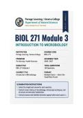
,BIOL 271 Module 3 Exam | Microbiology
w/Lab Q&A | Portage Learning
1. The ability of a microscope to distinguish two close objects as separate is
called:
A) Resolution
B) Magnification
C) Contrast
D) Refraction
Correct Answer: A) Resolution
Rationale: Resolution is the minimum distance between two points that can still
be seen as distinct. Magnification enlarges the image; contrast is the difference
in light intensity; refraction bends light but does not define resolving power.
2. Which unit of measurement is equal to one‑millionth of a meter and
commonly used for microbes?
A) Nanometer
B) Millimeter
C) Centimeter
D) Micrometer
Correct Answer: D) Micrometer
,Rationale: A micrometer (µm) is 10⁻⁶ m, suitable for measuring bacteria
(typically 1‑10 µm). A nanometer is 10⁻⁹ m, used for viruses and subcellular
structures.
3. The total magnification of a microscope is calculated by multiplying the
objective lens power by the:
A) Condenser power
B) Ocular lens power
C) Diaphragm setting
D) Stage adjustment
Correct Answer: B) Ocular lens power
Rationale: Total magnification = objective magnification × ocular magnification.
The most common ocular lens is 10×. The condenser and diaphragm control
light, not magnification.
4. The most common magnification power of the ocular lens in a standard light
microscope is:
A) 5×
B) 40×
C) 10×
D) 100×
Correct Answer: C) 10×
, Rationale: Ocular lenses typically magnify 10×. Combined with a 100× objective,
total magnification is 1000×. 5× is less common; 40× and 100× are objective
lenses.
5. The simplest form of light microscope, where light passes through the
specimen from below, is the:
A) Phase contrast microscope
B) Dark field microscope
C) Fluorescence microscope
D) Brightfield microscope
Correct Answer: D) Brightfield microscope
Rationale: Brightfield microscopy is the most basic, using transmitted light.
Phase contrast enhances unstained cells; dark field shows bright objects on a
dark background; fluorescence uses UV light.
6. A phase contrast microscope is especially useful for:
A) Viewing live, unstained cells by amplifying density differences
B) Observing fluorescently labeled antibodies
C) Scanning the surface of a specimen
D) Viewing internal structures at high resolution
Correct Answer: A) Viewing live, unstained cells by amplifying density
differences
w/Lab Q&A | Portage Learning
1. The ability of a microscope to distinguish two close objects as separate is
called:
A) Resolution
B) Magnification
C) Contrast
D) Refraction
Correct Answer: A) Resolution
Rationale: Resolution is the minimum distance between two points that can still
be seen as distinct. Magnification enlarges the image; contrast is the difference
in light intensity; refraction bends light but does not define resolving power.
2. Which unit of measurement is equal to one‑millionth of a meter and
commonly used for microbes?
A) Nanometer
B) Millimeter
C) Centimeter
D) Micrometer
Correct Answer: D) Micrometer
,Rationale: A micrometer (µm) is 10⁻⁶ m, suitable for measuring bacteria
(typically 1‑10 µm). A nanometer is 10⁻⁹ m, used for viruses and subcellular
structures.
3. The total magnification of a microscope is calculated by multiplying the
objective lens power by the:
A) Condenser power
B) Ocular lens power
C) Diaphragm setting
D) Stage adjustment
Correct Answer: B) Ocular lens power
Rationale: Total magnification = objective magnification × ocular magnification.
The most common ocular lens is 10×. The condenser and diaphragm control
light, not magnification.
4. The most common magnification power of the ocular lens in a standard light
microscope is:
A) 5×
B) 40×
C) 10×
D) 100×
Correct Answer: C) 10×
, Rationale: Ocular lenses typically magnify 10×. Combined with a 100× objective,
total magnification is 1000×. 5× is less common; 40× and 100× are objective
lenses.
5. The simplest form of light microscope, where light passes through the
specimen from below, is the:
A) Phase contrast microscope
B) Dark field microscope
C) Fluorescence microscope
D) Brightfield microscope
Correct Answer: D) Brightfield microscope
Rationale: Brightfield microscopy is the most basic, using transmitted light.
Phase contrast enhances unstained cells; dark field shows bright objects on a
dark background; fluorescence uses UV light.
6. A phase contrast microscope is especially useful for:
A) Viewing live, unstained cells by amplifying density differences
B) Observing fluorescently labeled antibodies
C) Scanning the surface of a specimen
D) Viewing internal structures at high resolution
Correct Answer: A) Viewing live, unstained cells by amplifying density
differences


