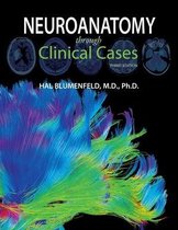
TEST BANK – NEUROANATOMY THROUGH CLINICAL CASES 3RD EDITION BY
BLUMENFELD
, Table of Contents
Chapter 1: Neuroanatomical Terminology
Chapter 2: Neural Cells and Their Response to Disease
Chapter 3: Peripheral Nervous System
Chapter 4: Spinal Cord
Chapter 5: Brain and Environs: Cranium, Ventricles, and Meninges
Chapter 6: Corticospinal Tract and Other Motor Pathways
Chapter 7: Somatosensory Pathways
Chapter 8: Spinal Nerve Roots
Chapter 9: Major Plexuses and Peripheral Nerves
Chapter 10: Cerebral Hemispheres and Vascular Supply
Chapter 11: Visual System
Chapter 12: Brainstem I: External Anatomy and Syndromes
Chapter 13: Brainstem II: Eye Movements and Pupillary Control
Chapter 14: Brainstem III: Internal Structures and Vascular Supply
Chapter 15: Cerebellum
Chapter 16: Basal Ganglia
Chapter 17: Pituitary and Hypothalamus
Chapter 18: Limbic System: Homeostasis, Olfaction, Memory and Emotion
Chapter 19: Higher-Order Cerebral Function
Answer Key with Rationales
• Detailed Explanations for Every Question
• Clinical Correlations
• High-Yield Neuroanatomy Review
• Board-Style Learning Points
, Chapter 1: Introduction to Clinical Case Presentations
Challenge 1
During an outpatient consultation, a patient suddenly develops fluent but meaningless speech and is
unable to follow simple verbal commands. Motor strength, coordination, and sensation remain intact.
Before ordering additional investigations, which principle should guide the clinician's diagnostic reasoning?
A. Determine the most likely disease before lesion localization.
B. Delay localization until MRI findings are available.
C. Prioritize laboratory investigations over bedside examination.
D. Localize the lesion before determining the underlying disease process.
Correct Answer: D. Localize the lesion before determining the underlying disease process.
Rationale
Neurologic diagnosis begins with anatomical localization. Identifying where the lesion is located allows
clinicians to narrow the differential diagnosis before determining the specific pathology. This localization-first
approach is a defining feature of clinical neurology and underpins effective diagnostic reasoning.
Why the Other Options Are Less Appropriate
A. Diagnosis without localization often results in an unnecessarily broad differential.
B. MRI confirms clinical suspicion but should not replace bedside localization.
C. Laboratory investigations rarely establish the anatomical location of a focal neurologic lesion.
Clinical Pearl
Always answer "Where is the lesion?" before asking "What caused it?"
Board Exam Tip
Board examinations frequently test localization before diagnosis; focus on the neuroanatomical pattern
rather than the disease label.
Challenge 2
While presenting a new referral, a resident concludes that the patient has multiple sclerosis after
hearing only that numbness developed over several days. Which missing component of the clinical
presentation most limits the accuracy of this conclusion?
,A. A detailed description of symptom chronology and progression.
B. Family history of hypertension.
C. Blood group determination.
D. Occupation and educational background.
Correct Answer: A. A detailed description of symptom chronology and progression.
Rationale
The onset and progression of neurologic symptoms are essential for differentiating vascular, inflammatory,
infectious, neoplastic, degenerative, and metabolic disorders. A precise timeline strengthens localization and
guides the diagnostic process.
Why the Other Options Are Less Appropriate
B. Hypertension is a risk factor but does not replace a complete neurologic history.
C. Blood type has little value in lesion localization.
D. Social history is helpful but secondary to symptom evolution.
Clinical Pearl
The tempo of disease is often as informative as the physical examination.
Board Exam Tip
Always correlate lesion location with symptom progression before choosing a diagnosis.
Challenge 3
MRI reveals a small lesion within the posterior limb of the internal capsule. The patient has dense
contralateral weakness but normal cognition, language, vision, and sensation. Which principle best explains
why a relatively small lesion produced profound disability?
A. The lesion interrupts diffuse cortical association pathways.
B. The internal capsule contains densely packed descending motor fibers.
C. Small subcortical lesions primarily affect memory circuits.
D. White matter lesions rarely correlate with clinical severity.
Correct Answer: B. The internal capsule contains densely packed descending motor fibers.
Rationale
,The posterior limb of the internal capsule contains highly concentrated corticospinal fibers. Even a small lesion
can interrupt numerous descending motor pathways simultaneously, producing severe contralateral motor
deficits despite minimal radiologic size.
Why the Other Options Are Less Appropriate
A. Association pathways are primarily involved in higher cortical function.
C. Memory circuits are not the principal structures affected.
D. Clinical severity often reflects lesion location more than lesion size.
Clinical Pearl
In neurology, strategic lesion location frequently outweighs lesion volume.
Board Exam Tip
Small lesions affecting compact white matter tracts often produce disproportionately severe deficits.
Challenge 4
Neurologic examination identifies ipsilateral facial weakness with contralateral limb weakness. Before
considering the specific disease, where should the lesion be localized?
A. Cerebellar hemisphere.
B. Cervical spinal cord.
C. Brainstem.
D. Primary motor cortex.
Correct Answer: C. Brainstem.
Rationale
Crossed neurologic findings—cranial nerve deficits on one side with long-tract motor or sensory deficits on the
opposite side—are classic indicators of a brainstem lesion. This localization is one of the most reliable principles
in clinical neurology.
Why the Other Options Are Less Appropriate
A. Cerebellar lesions produce ipsilateral coordination deficits rather than crossed findings.
B. The spinal cord does not contain cranial nerve nuclei.
D. Cortical lesions typically produce contralateral deficits without ipsilateral cranial nerve involvement.
Clinical Pearl
Crossed findings almost always indicate a brainstem lesion until proven otherwise.
, Board Exam Tip
Whenever cranial nerve abnormalities accompany contralateral body deficits, localize to the brainstem first.
Challenge 5
During visual field testing, a patient consistently fails to detect objects in the right visual field of both
eyes. Which clinical reasoning step should immediately follow identifying this deficit?
A. Determine the exact neuroanatomical location along the visual pathway.
B. Diagnose occipital stroke without further assessment.
C. Request lumbar puncture before examination.
D. Assume an ophthalmologic disorder is responsible.
Correct Answer: A. Determine the exact neuroanatomical location along the visual pathway.
Rationale
Visual field defects localize to specific portions of the visual pathway. Accurate localization differentiates
retinal, optic nerve, optic tract, optic radiation, and occipital lesions before determining the underlying disease.
Why the Other Options Are Less Appropriate
B. Several lesions can produce similar field defects.
C. Lumbar puncture is not the next logical diagnostic step.
D. Homonymous field defects usually indicate retrochiasmal pathology.
Clinical Pearl
The pattern of visual loss often identifies the lesion before imaging does.
Board Exam Tip
Always map visual field deficits anatomically before considering etiology.
Challenge 6
Moments after recovering from a generalized seizure, a patient exhibits isolated weakness of the left
arm that resolves completely within 30 minutes. Which explanation best accounts for this transient deficit?
A. Acute spinal cord compression.
B. Todd paralysis following focal cortical dysfunction.
C. Peripheral nerve transection.
,D. Progressive motor neuron disease.
Correct Answer: B. Todd paralysis following focal cortical dysfunction.
Rationale
Transient postictal weakness, known as Todd paralysis, occurs after focal cortical seizure activity and resolves
spontaneously. Recognizing this phenomenon prevents unnecessary misdiagnosis of acute stroke while
ensuring appropriate evaluation.
Why the Other Options Are Less Appropriate
A. Spinal cord compression produces persistent deficits.
C. Peripheral nerve injury does not resolve rapidly.
D. Motor neuron disease progresses gradually rather than resolving spontaneously.
Clinical Pearl
Temporary focal weakness after a seizure should always raise suspicion for Todd paralysis.
Board Exam Tip
Differentiate postictal deficits from acute stroke using history, examination, and symptom evolution.
Challenge 7
Genetic testing confirms a hereditary neurologic disorder in several members of one family. During
evaluation of an asymptomatic relative, which component of the clinical presentation remains most
important despite the known genetic diagnosis?
A. Precise neuroanatomical localization.
B. Blood glucose measurement alone.
C. Routine chest radiography.
D. Serum electrolyte screening before examination.
Correct Answer: A. Precise neuroanatomical localization.
Rationale
Even when the underlying disease is known, localization remains essential because it defines disease severity,
explains symptoms, guides management, and predicts functional impairment.
Why the Other Options Are Less Appropriate
B. Glucose testing cannot localize neurologic dysfunction.
C. Chest radiography has limited relevance here.
,D. Electrolytes complement but do not replace neurologic assessment.
Clinical Pearl
Diagnosis identifies the disease; localization explains the patient's symptoms.
Board Exam Tip
Never abandon localization simply because the diagnosis appears obvious.
Challenge 8
Following transfer to the neuroscience ICU, a patient opens the eyes only to painful stimulation,
produces incomprehensible sounds, and withdraws from pain. Which assessment principle is being applied
to communicate neurologic status objectively?
A. Functional Independence Measure.
B. National Institutes of Health Stroke Scale.
C. Glasgow Coma Scale.
D. Modified Rankin Scale.
Correct Answer: C. Glasgow Coma Scale.
Rationale
The Glasgow Coma Scale objectively evaluates eye opening, verbal response, and motor response, providing a
standardized method for assessing level of consciousness and monitoring changes over time.
Why the Other Options Are Less Appropriate
A. Measures functional independence during rehabilitation.
B. Assesses stroke-related neurologic deficits.
D. Measures long-term disability after stroke.
Clinical Pearl
Objective neurologic scales improve communication between clinicians and facilitate serial assessments.
Board Exam Tip
Know the components of commonly used neurologic scoring systems and when each is most appropriate.
Challenge 9
, While reviewing a complex neurologic referral, the consultant notes that every reported symptom can
be explained by a single lesion. Which diagnostic principle is most consistent with this observation?
A. Multiple simultaneous lesions should always be assumed.
B. The simplest anatomical explanation that accounts for all findings should be considered first.
C. Rare diseases should always be prioritized.
D. Laboratory abnormalities outweigh examination findings.
Correct Answer: B. The simplest anatomical explanation that accounts for all findings should be
considered first.
Rationale
Whenever possible, neurologists seek a single lesion capable of explaining the complete clinical presentation.
This approach promotes efficient localization and avoids unnecessary diagnostic complexity.
Why the Other Options Are Less Appropriate
A. Multiple lesions are considered only when one lesion cannot explain the findings.
C. Common explanations should generally be evaluated before rare conditions.
D. Clinical localization remains central to neurologic diagnosis.
Clinical Pearl
A single well-localized lesion is often more likely than multiple unrelated pathologies.
Board Exam Tip
Look for one lesion that explains every deficit before proposing multiple disease processes.
Challenge 10
During the final review of a new consultation, the attending neurologist reminds trainees that an
excellent clinical case presentation should lead listeners toward the diagnosis before investigations are
revealed. Which feature most consistently distinguishes an outstanding neurologic presentation?
A. Listing every laboratory value obtained.
B. Presenting imaging findings before the history.
C. Emphasizing rare disorders early in the discussion.
D. Organizing the history and examination to support logical neuroanatomical localization.
Correct Answer: D. Organizing the history and examination to support logical neuroanatomical
localization.
, Rationale
The hallmark of an effective neurologic case presentation is a logical sequence that allows listeners to localize
the lesion before considering etiology. A structured presentation improves communication, diagnostic accuracy,
and clinical decision-making.
Why the Other Options Are Less Appropriate
A. Laboratory values should support—not dominate—the presentation.
B. History and examination should precede interpretation of investigations.
C. Common conditions should generally be considered before rare disorders.
Clinical Pearl
A well-constructed neurologic presentation tells a diagnostic story that naturally progresses from history to
localization to diagnosis.
Board Exam Tip
Examiners often assess your clinical reasoning through the organization of your presentation as much as
through the final diagnosis.
Challenge 11
Air ambulance transfers a patient after sudden collapse at work. Neurologic examination demonstrates
complete left facial paralysis involving the forehead, left-sided hearing loss, absent corneal reflex on the left,
and contralateral loss of pain and temperature sensation over the body. Which diagnostic approach should
be prioritized before identifying the underlying disease?
A. Determine the vascular territory involved before localization.
B. Begin empirical antimicrobial therapy immediately.
C. Localize the lesion to a specific neuroanatomical region.
D. Request cerebrospinal fluid analysis before bedside assessment.
Correct Answer: C. Localize the lesion to a specific neuroanatomical region.
Rationale
The combination of ipsilateral cranial nerve deficits and contralateral sensory loss strongly suggests a
brainstem lesion. Before determining whether the cause is ischemic, inflammatory, neoplastic, or infectious, the
clinician should first localize the lesion anatomically. Accurate localization significantly narrows the differential
diagnosis and guides targeted investigations.
Why the Other Options Are Less Appropriate
BLUMENFELD
, Table of Contents
Chapter 1: Neuroanatomical Terminology
Chapter 2: Neural Cells and Their Response to Disease
Chapter 3: Peripheral Nervous System
Chapter 4: Spinal Cord
Chapter 5: Brain and Environs: Cranium, Ventricles, and Meninges
Chapter 6: Corticospinal Tract and Other Motor Pathways
Chapter 7: Somatosensory Pathways
Chapter 8: Spinal Nerve Roots
Chapter 9: Major Plexuses and Peripheral Nerves
Chapter 10: Cerebral Hemispheres and Vascular Supply
Chapter 11: Visual System
Chapter 12: Brainstem I: External Anatomy and Syndromes
Chapter 13: Brainstem II: Eye Movements and Pupillary Control
Chapter 14: Brainstem III: Internal Structures and Vascular Supply
Chapter 15: Cerebellum
Chapter 16: Basal Ganglia
Chapter 17: Pituitary and Hypothalamus
Chapter 18: Limbic System: Homeostasis, Olfaction, Memory and Emotion
Chapter 19: Higher-Order Cerebral Function
Answer Key with Rationales
• Detailed Explanations for Every Question
• Clinical Correlations
• High-Yield Neuroanatomy Review
• Board-Style Learning Points
, Chapter 1: Introduction to Clinical Case Presentations
Challenge 1
During an outpatient consultation, a patient suddenly develops fluent but meaningless speech and is
unable to follow simple verbal commands. Motor strength, coordination, and sensation remain intact.
Before ordering additional investigations, which principle should guide the clinician's diagnostic reasoning?
A. Determine the most likely disease before lesion localization.
B. Delay localization until MRI findings are available.
C. Prioritize laboratory investigations over bedside examination.
D. Localize the lesion before determining the underlying disease process.
Correct Answer: D. Localize the lesion before determining the underlying disease process.
Rationale
Neurologic diagnosis begins with anatomical localization. Identifying where the lesion is located allows
clinicians to narrow the differential diagnosis before determining the specific pathology. This localization-first
approach is a defining feature of clinical neurology and underpins effective diagnostic reasoning.
Why the Other Options Are Less Appropriate
A. Diagnosis without localization often results in an unnecessarily broad differential.
B. MRI confirms clinical suspicion but should not replace bedside localization.
C. Laboratory investigations rarely establish the anatomical location of a focal neurologic lesion.
Clinical Pearl
Always answer "Where is the lesion?" before asking "What caused it?"
Board Exam Tip
Board examinations frequently test localization before diagnosis; focus on the neuroanatomical pattern
rather than the disease label.
Challenge 2
While presenting a new referral, a resident concludes that the patient has multiple sclerosis after
hearing only that numbness developed over several days. Which missing component of the clinical
presentation most limits the accuracy of this conclusion?
,A. A detailed description of symptom chronology and progression.
B. Family history of hypertension.
C. Blood group determination.
D. Occupation and educational background.
Correct Answer: A. A detailed description of symptom chronology and progression.
Rationale
The onset and progression of neurologic symptoms are essential for differentiating vascular, inflammatory,
infectious, neoplastic, degenerative, and metabolic disorders. A precise timeline strengthens localization and
guides the diagnostic process.
Why the Other Options Are Less Appropriate
B. Hypertension is a risk factor but does not replace a complete neurologic history.
C. Blood type has little value in lesion localization.
D. Social history is helpful but secondary to symptom evolution.
Clinical Pearl
The tempo of disease is often as informative as the physical examination.
Board Exam Tip
Always correlate lesion location with symptom progression before choosing a diagnosis.
Challenge 3
MRI reveals a small lesion within the posterior limb of the internal capsule. The patient has dense
contralateral weakness but normal cognition, language, vision, and sensation. Which principle best explains
why a relatively small lesion produced profound disability?
A. The lesion interrupts diffuse cortical association pathways.
B. The internal capsule contains densely packed descending motor fibers.
C. Small subcortical lesions primarily affect memory circuits.
D. White matter lesions rarely correlate with clinical severity.
Correct Answer: B. The internal capsule contains densely packed descending motor fibers.
Rationale
,The posterior limb of the internal capsule contains highly concentrated corticospinal fibers. Even a small lesion
can interrupt numerous descending motor pathways simultaneously, producing severe contralateral motor
deficits despite minimal radiologic size.
Why the Other Options Are Less Appropriate
A. Association pathways are primarily involved in higher cortical function.
C. Memory circuits are not the principal structures affected.
D. Clinical severity often reflects lesion location more than lesion size.
Clinical Pearl
In neurology, strategic lesion location frequently outweighs lesion volume.
Board Exam Tip
Small lesions affecting compact white matter tracts often produce disproportionately severe deficits.
Challenge 4
Neurologic examination identifies ipsilateral facial weakness with contralateral limb weakness. Before
considering the specific disease, where should the lesion be localized?
A. Cerebellar hemisphere.
B. Cervical spinal cord.
C. Brainstem.
D. Primary motor cortex.
Correct Answer: C. Brainstem.
Rationale
Crossed neurologic findings—cranial nerve deficits on one side with long-tract motor or sensory deficits on the
opposite side—are classic indicators of a brainstem lesion. This localization is one of the most reliable principles
in clinical neurology.
Why the Other Options Are Less Appropriate
A. Cerebellar lesions produce ipsilateral coordination deficits rather than crossed findings.
B. The spinal cord does not contain cranial nerve nuclei.
D. Cortical lesions typically produce contralateral deficits without ipsilateral cranial nerve involvement.
Clinical Pearl
Crossed findings almost always indicate a brainstem lesion until proven otherwise.
, Board Exam Tip
Whenever cranial nerve abnormalities accompany contralateral body deficits, localize to the brainstem first.
Challenge 5
During visual field testing, a patient consistently fails to detect objects in the right visual field of both
eyes. Which clinical reasoning step should immediately follow identifying this deficit?
A. Determine the exact neuroanatomical location along the visual pathway.
B. Diagnose occipital stroke without further assessment.
C. Request lumbar puncture before examination.
D. Assume an ophthalmologic disorder is responsible.
Correct Answer: A. Determine the exact neuroanatomical location along the visual pathway.
Rationale
Visual field defects localize to specific portions of the visual pathway. Accurate localization differentiates
retinal, optic nerve, optic tract, optic radiation, and occipital lesions before determining the underlying disease.
Why the Other Options Are Less Appropriate
B. Several lesions can produce similar field defects.
C. Lumbar puncture is not the next logical diagnostic step.
D. Homonymous field defects usually indicate retrochiasmal pathology.
Clinical Pearl
The pattern of visual loss often identifies the lesion before imaging does.
Board Exam Tip
Always map visual field deficits anatomically before considering etiology.
Challenge 6
Moments after recovering from a generalized seizure, a patient exhibits isolated weakness of the left
arm that resolves completely within 30 minutes. Which explanation best accounts for this transient deficit?
A. Acute spinal cord compression.
B. Todd paralysis following focal cortical dysfunction.
C. Peripheral nerve transection.
,D. Progressive motor neuron disease.
Correct Answer: B. Todd paralysis following focal cortical dysfunction.
Rationale
Transient postictal weakness, known as Todd paralysis, occurs after focal cortical seizure activity and resolves
spontaneously. Recognizing this phenomenon prevents unnecessary misdiagnosis of acute stroke while
ensuring appropriate evaluation.
Why the Other Options Are Less Appropriate
A. Spinal cord compression produces persistent deficits.
C. Peripheral nerve injury does not resolve rapidly.
D. Motor neuron disease progresses gradually rather than resolving spontaneously.
Clinical Pearl
Temporary focal weakness after a seizure should always raise suspicion for Todd paralysis.
Board Exam Tip
Differentiate postictal deficits from acute stroke using history, examination, and symptom evolution.
Challenge 7
Genetic testing confirms a hereditary neurologic disorder in several members of one family. During
evaluation of an asymptomatic relative, which component of the clinical presentation remains most
important despite the known genetic diagnosis?
A. Precise neuroanatomical localization.
B. Blood glucose measurement alone.
C. Routine chest radiography.
D. Serum electrolyte screening before examination.
Correct Answer: A. Precise neuroanatomical localization.
Rationale
Even when the underlying disease is known, localization remains essential because it defines disease severity,
explains symptoms, guides management, and predicts functional impairment.
Why the Other Options Are Less Appropriate
B. Glucose testing cannot localize neurologic dysfunction.
C. Chest radiography has limited relevance here.
,D. Electrolytes complement but do not replace neurologic assessment.
Clinical Pearl
Diagnosis identifies the disease; localization explains the patient's symptoms.
Board Exam Tip
Never abandon localization simply because the diagnosis appears obvious.
Challenge 8
Following transfer to the neuroscience ICU, a patient opens the eyes only to painful stimulation,
produces incomprehensible sounds, and withdraws from pain. Which assessment principle is being applied
to communicate neurologic status objectively?
A. Functional Independence Measure.
B. National Institutes of Health Stroke Scale.
C. Glasgow Coma Scale.
D. Modified Rankin Scale.
Correct Answer: C. Glasgow Coma Scale.
Rationale
The Glasgow Coma Scale objectively evaluates eye opening, verbal response, and motor response, providing a
standardized method for assessing level of consciousness and monitoring changes over time.
Why the Other Options Are Less Appropriate
A. Measures functional independence during rehabilitation.
B. Assesses stroke-related neurologic deficits.
D. Measures long-term disability after stroke.
Clinical Pearl
Objective neurologic scales improve communication between clinicians and facilitate serial assessments.
Board Exam Tip
Know the components of commonly used neurologic scoring systems and when each is most appropriate.
Challenge 9
, While reviewing a complex neurologic referral, the consultant notes that every reported symptom can
be explained by a single lesion. Which diagnostic principle is most consistent with this observation?
A. Multiple simultaneous lesions should always be assumed.
B. The simplest anatomical explanation that accounts for all findings should be considered first.
C. Rare diseases should always be prioritized.
D. Laboratory abnormalities outweigh examination findings.
Correct Answer: B. The simplest anatomical explanation that accounts for all findings should be
considered first.
Rationale
Whenever possible, neurologists seek a single lesion capable of explaining the complete clinical presentation.
This approach promotes efficient localization and avoids unnecessary diagnostic complexity.
Why the Other Options Are Less Appropriate
A. Multiple lesions are considered only when one lesion cannot explain the findings.
C. Common explanations should generally be evaluated before rare conditions.
D. Clinical localization remains central to neurologic diagnosis.
Clinical Pearl
A single well-localized lesion is often more likely than multiple unrelated pathologies.
Board Exam Tip
Look for one lesion that explains every deficit before proposing multiple disease processes.
Challenge 10
During the final review of a new consultation, the attending neurologist reminds trainees that an
excellent clinical case presentation should lead listeners toward the diagnosis before investigations are
revealed. Which feature most consistently distinguishes an outstanding neurologic presentation?
A. Listing every laboratory value obtained.
B. Presenting imaging findings before the history.
C. Emphasizing rare disorders early in the discussion.
D. Organizing the history and examination to support logical neuroanatomical localization.
Correct Answer: D. Organizing the history and examination to support logical neuroanatomical
localization.
, Rationale
The hallmark of an effective neurologic case presentation is a logical sequence that allows listeners to localize
the lesion before considering etiology. A structured presentation improves communication, diagnostic accuracy,
and clinical decision-making.
Why the Other Options Are Less Appropriate
A. Laboratory values should support—not dominate—the presentation.
B. History and examination should precede interpretation of investigations.
C. Common conditions should generally be considered before rare disorders.
Clinical Pearl
A well-constructed neurologic presentation tells a diagnostic story that naturally progresses from history to
localization to diagnosis.
Board Exam Tip
Examiners often assess your clinical reasoning through the organization of your presentation as much as
through the final diagnosis.
Challenge 11
Air ambulance transfers a patient after sudden collapse at work. Neurologic examination demonstrates
complete left facial paralysis involving the forehead, left-sided hearing loss, absent corneal reflex on the left,
and contralateral loss of pain and temperature sensation over the body. Which diagnostic approach should
be prioritized before identifying the underlying disease?
A. Determine the vascular territory involved before localization.
B. Begin empirical antimicrobial therapy immediately.
C. Localize the lesion to a specific neuroanatomical region.
D. Request cerebrospinal fluid analysis before bedside assessment.
Correct Answer: C. Localize the lesion to a specific neuroanatomical region.
Rationale
The combination of ipsilateral cranial nerve deficits and contralateral sensory loss strongly suggests a
brainstem lesion. Before determining whether the cause is ischemic, inflammatory, neoplastic, or infectious, the
clinician should first localize the lesion anatomically. Accurate localization significantly narrows the differential
diagnosis and guides targeted investigations.
Why the Other Options Are Less Appropriate