

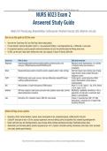
NURS 6023 Exam 2 Answered Study Guide | Respiratory, Breast, CV/PV, EKG, Abdomen, Labs
NURS 6023 Exam 2
Answered Study Guide
Weeks 5-8: Thorax/Lungs, Breasts/Axillae, Cardiovascular, Peripheral Vascular, EKG, Abdomen, and Labs
How to use this guide to ACE the exam
Start with the "Exam Power Grid" and "Red Flags" before reading details.
For each disorder, memorize the pattern: what it is -> key assessment findings -> best diagnostic/lab clue -> differential -> exam pearl.
For assessment questions, practice saying the normal documentation out loud, then add what abnormal findings would mean.
For EKG, use the same 5-step rhythm method every time: rate, regularity, P waves, PR interval, QRS width.
Exam area What to know High-yield remember
Respiratory Inspection/palpation/percussion/auscultation patterns; abnormal sounds; vocal Resonance normal; hyperresonance = air; dullness
resonance; COPD/asthma/pneumonia/PTX/PE/effusion/TB. = fluid/solid. Positive vocal resonance =
consolidation.
Breast Mass/pain/discharge questions; inspection positions; palpation pattern; nodes; red flags. Hard fixed irregular mass, dimpling, peau d'orange,
nipple retraction, bloody unilateral discharge =
malignancy concern.
CV/PV PMI/JVP/carotids; heart sounds; murmurs; chest pain differentials; edema/DVT/venous S3 after age 40 = systolic dysfunction; MR/AS
insufficiency/PAD/carotid stenosis. systolic, MS/AR diastolic; arterial disease is
cool/dry/pulseless.
EKG NSR parameters; 13 rhythm disturbances; STEMI locations. Inferior = II, III, aVF. Lateral = I, aVL, V5-V6. Anterior
= V3-V4. Septal = V1-V2.
Abdomen IAPP order; inspection, bowel sounds, percussion notes, palpation, special signs, common RUQ/Murphy = gallbladder. Periumbilical -> RLQ =
abdominal/GU/endocrine disorders. appendicitis. LLQ older adult = diverticulitis. Back
radiation = pancreatitis/AAA.
Labs Electrolytes, LFTs, cholesterol, troponin, BNP, CBC, renal markers. Troponin = myocardial injury. BNP = HF. K/Mg
abnormalities = arrhythmia risk. LFT pattern helps
liver/biliary differential.
Global red flags across systems
Respiratory: stridor, tracheal deviation, cyanosis, severe new dyspnea at rest, unexplained hypoxia, sudden pleuritic chest pain.
Cardiac/PV: chest pain at rest or >20 min, syncope, hypotension, new neuro deficits, painful cold pulseless limb, unilateral leg swelling/warmth.
Breast: hard fixed mass, skin dimpling/retraction, peau d'orange, bloody unilateral spontaneous discharge, fixed/matted axillary nodes.
Abdomen/GU: pain that wakes patient, persistent progressive pain >6 hr, migration, intractable vomiting, hematemesis, black stools, shock, decreased
urine output, painless gross hematuria.
High-yield review from uploaded lecture/study-guide sources. Verify nuance with assigned Bates/Goolsby readings.
, NURS 6023 Exam 2 Answered Study Guide | Respiratory, Breast, CV/PV, EKG, Abdomen, Labs
Week 5 - Thorax and Lungs: Completed Student Notes
Key point from study guide Completed student notes
Appropriate HPI questions for a Use OLD CARTS plus respiratory-specific questions. Ask onset/duration; cough dry vs productive; sputum amount, color, odor;
respiratory chief complaint hemoptysis and whether blood is truly from lungs vs mouth/nose; dyspnea at rest/exertion and change from baseline; wheeze; chest
pain and whether pleuritic; fever/chills/night sweats/weight loss; sick contacts; travel/TB exposure; occupational or chemical
exposures; smoking/vaping history and pack-years; asthma/COPD/pneumonia history; allergies; current medications including ACE
inhibitors; immunization status; and prior CXR, CT, PFT, or inhaler use.
Inspecting the chest: size, shape, Expose the chest appropriately and compare side-to-side. Expected: chest symmetric, AP diameter less than transverse diameter, no
symmetry, landmarks retractions, no accessory muscle use, no visible deformity, and respirations unlabored. Abnormal: barrel chest/increased AP diameter
with COPD/emphysema; pectus excavatum or carinatum; kyphosis/scoliosis; unilateral lag with pneumothorax, effusion, rib fracture, or
severe pneumonia; retractions/nasal flaring/pursed-lip breathing with increased work of breathing. Landmarks: lung apices are 2-4 cm
above the clavicles anteriorly and around C7 posteriorly; bases are around the 6th rib at the midclavicular line anteriorly and T10
posteriorly; right lung has 3 lobes, left lung has 2 lobes; right middle lobe is heard best anteriorly.
Assessing rate and quality of Count respirations while the patient is at rest. Expected adult rate is about 12-20/min, regular, quiet, and unlabored with symmetric
respirations chest rise. Document rate, rhythm/pattern, depth, effort, and accessory muscle use. Abnormal: tachypnea, bradypnea, apnea, shallow
respirations, labored breathing, nasal flaring, retractions, pursed-lip breathing, tripod posture, Cheyne-Stokes, Kussmaul, or Biot
respirations. Documentation example: 'Respirations 18/min, regular and unlabored, symmetric chest expansion, no accessory muscle
use.'
Peripheral assessment: lips, nails, skin Expected: lips and mucous membranes pink, nail beds pink, capillary refill usually less than 2 seconds, no cyanosis, no clubbing.
color Abnormal: central cyanosis of lips/mucous membranes suggests hypoxemia and is a late sign; peripheral cyanosis may suggest poor
peripheral perfusion; clubbing suggests chronic hypoxia or chronic cardiopulmonary disease; pallor may suggest anemia; diaphoresis
can occur with respiratory distress, cardiac ischemia, or infection. Document exactly what is seen.
Palpating chest and trachea; tactile Palpate the chest wall for tenderness, masses, crepitus, and symmetry. Trachea should be midline. Assess thoracic expansion by
fremitus and thoracic expansion placing thumbs near T9/T10 posteriorly and watching for equal thumb separation as the patient inhales. Tactile fremitus is palpable
vibration transmitted through the chest wall when the patient speaks, usually '99.' Expected fremitus is symmetric. Increased fremitus
suggests denser lung tissue such as consolidation/pneumonia. Decreased fremitus suggests blocked transmission such as pleural
effusion, pneumothorax, emphysema/COPD, or bronchial obstruction.
Percussing the chest Percuss side-to-side in a ladder pattern over comparable lung areas. Expected lung note is resonance. Hyperresonance means too
much air, such as COPD/emphysema or pneumothorax. Dullness means fluid or solid tissue, such as pneumonia/consolidation, pleural
effusion, tumor, or atelectasis. Flatness is expected over bone or muscle. Tympany over the lung is abnormal and can suggest
pneumothorax or a large air space.
Measuring diaphragmatic excursion Ask the patient to exhale fully and hold; percuss down the scapular line from resonance to dullness and mark. Then ask the patient to
inhale deeply and hold; percuss down again until dullness and mark. Measure the distance between marks. Expected excursion is
generally about 3-5 cm and should be symmetric. Decreased or asymmetric excursion can occur with COPD hyperinflation,
diaphragmatic paralysis, pleural effusion, atelectasis, or severe lower-lobe disease.
Normal breath sounds: types, location, Vesicular: soft, low-pitched, heard over most peripheral lung fields; inspiration is longer than expiration. Bronchovesicular: medium
description pitch/intensity, heard over major bronchi - 1st/2nd interspaces anteriorly and between scapulae posteriorly; inspiration and expiration
are about equal. Bronchial: louder, higher-pitched, heard over the manubrium/large airways; expiration is longer than inspiration.
Tracheal sounds over the trachea are very loud/high-pitched and are expected only over the trachea.
Abnormal breath sounds and likely Crackles/rales: discontinuous popping; air moving through fluid or opening collapsed alveoli; think pneumonia, heart failure,
differentials pulmonary edema, fibrosis. Wheezes: high-pitched musical sounds; narrowed airways; think asthma, COPD, bronchitis. Rhonchi: low-
pitched snoring/gurgling that may clear with cough; secretions in larger airways; think bronchitis or retained secretions. Stridor: harsh,
high-pitched, loudest over the neck; upper airway obstruction; urgent. Pleural friction rub: grating/leathery sound with inspiration and
expiration; inflamed pleural surfaces; think pleurisy, pneumonia, pulmonary embolism, or pleural inflammation. Diminished/absent
sounds: poor air movement, effusion, pneumothorax, obstruction, or severe COPD.
Three types of vocal resonance Bronchophony: patient says '99'; abnormal if words sound louder/clearer instead of muffled. Egophony: patient says 'E'; abnormal if it
sounds like 'A.' Whispered pectoriloquy: patient whispers '99'; abnormal if whisper is louder/clearer. Positive vocal resonance suggests
consolidation, commonly pneumonia. Egophony can also occur at the upper level of a pleural effusion.
Documenting abnormal lung findings Document location, side, quality, and associated findings. Examples: 'Fine crackles noted in bilateral lower lobes, greater on right, no
wheezes.' 'Diffuse expiratory wheezes throughout posterior lung fields, prolonged expiratory phase.' 'Diminished breath sounds over
left lower posterior field with dullness to percussion and decreased tactile fremitus.' 'Trachea deviated right; absent breath sounds on
left; patient dyspneic' - urgent finding.
High-yield review from uploaded lecture/study-guide sources. Verify nuance with assigned Bates/Goolsby readings.
NURS 6023 Exam 2
Answered Study Guide
Weeks 5-8: Thorax/Lungs, Breasts/Axillae, Cardiovascular, Peripheral Vascular, EKG, Abdomen, and Labs
How to use this guide to ACE the exam
Start with the "Exam Power Grid" and "Red Flags" before reading details.
For each disorder, memorize the pattern: what it is -> key assessment findings -> best diagnostic/lab clue -> differential -> exam pearl.
For assessment questions, practice saying the normal documentation out loud, then add what abnormal findings would mean.
For EKG, use the same 5-step rhythm method every time: rate, regularity, P waves, PR interval, QRS width.
Exam area What to know High-yield remember
Respiratory Inspection/palpation/percussion/auscultation patterns; abnormal sounds; vocal Resonance normal; hyperresonance = air; dullness
resonance; COPD/asthma/pneumonia/PTX/PE/effusion/TB. = fluid/solid. Positive vocal resonance =
consolidation.
Breast Mass/pain/discharge questions; inspection positions; palpation pattern; nodes; red flags. Hard fixed irregular mass, dimpling, peau d'orange,
nipple retraction, bloody unilateral discharge =
malignancy concern.
CV/PV PMI/JVP/carotids; heart sounds; murmurs; chest pain differentials; edema/DVT/venous S3 after age 40 = systolic dysfunction; MR/AS
insufficiency/PAD/carotid stenosis. systolic, MS/AR diastolic; arterial disease is
cool/dry/pulseless.
EKG NSR parameters; 13 rhythm disturbances; STEMI locations. Inferior = II, III, aVF. Lateral = I, aVL, V5-V6. Anterior
= V3-V4. Septal = V1-V2.
Abdomen IAPP order; inspection, bowel sounds, percussion notes, palpation, special signs, common RUQ/Murphy = gallbladder. Periumbilical -> RLQ =
abdominal/GU/endocrine disorders. appendicitis. LLQ older adult = diverticulitis. Back
radiation = pancreatitis/AAA.
Labs Electrolytes, LFTs, cholesterol, troponin, BNP, CBC, renal markers. Troponin = myocardial injury. BNP = HF. K/Mg
abnormalities = arrhythmia risk. LFT pattern helps
liver/biliary differential.
Global red flags across systems
Respiratory: stridor, tracheal deviation, cyanosis, severe new dyspnea at rest, unexplained hypoxia, sudden pleuritic chest pain.
Cardiac/PV: chest pain at rest or >20 min, syncope, hypotension, new neuro deficits, painful cold pulseless limb, unilateral leg swelling/warmth.
Breast: hard fixed mass, skin dimpling/retraction, peau d'orange, bloody unilateral spontaneous discharge, fixed/matted axillary nodes.
Abdomen/GU: pain that wakes patient, persistent progressive pain >6 hr, migration, intractable vomiting, hematemesis, black stools, shock, decreased
urine output, painless gross hematuria.
High-yield review from uploaded lecture/study-guide sources. Verify nuance with assigned Bates/Goolsby readings.
, NURS 6023 Exam 2 Answered Study Guide | Respiratory, Breast, CV/PV, EKG, Abdomen, Labs
Week 5 - Thorax and Lungs: Completed Student Notes
Key point from study guide Completed student notes
Appropriate HPI questions for a Use OLD CARTS plus respiratory-specific questions. Ask onset/duration; cough dry vs productive; sputum amount, color, odor;
respiratory chief complaint hemoptysis and whether blood is truly from lungs vs mouth/nose; dyspnea at rest/exertion and change from baseline; wheeze; chest
pain and whether pleuritic; fever/chills/night sweats/weight loss; sick contacts; travel/TB exposure; occupational or chemical
exposures; smoking/vaping history and pack-years; asthma/COPD/pneumonia history; allergies; current medications including ACE
inhibitors; immunization status; and prior CXR, CT, PFT, or inhaler use.
Inspecting the chest: size, shape, Expose the chest appropriately and compare side-to-side. Expected: chest symmetric, AP diameter less than transverse diameter, no
symmetry, landmarks retractions, no accessory muscle use, no visible deformity, and respirations unlabored. Abnormal: barrel chest/increased AP diameter
with COPD/emphysema; pectus excavatum or carinatum; kyphosis/scoliosis; unilateral lag with pneumothorax, effusion, rib fracture, or
severe pneumonia; retractions/nasal flaring/pursed-lip breathing with increased work of breathing. Landmarks: lung apices are 2-4 cm
above the clavicles anteriorly and around C7 posteriorly; bases are around the 6th rib at the midclavicular line anteriorly and T10
posteriorly; right lung has 3 lobes, left lung has 2 lobes; right middle lobe is heard best anteriorly.
Assessing rate and quality of Count respirations while the patient is at rest. Expected adult rate is about 12-20/min, regular, quiet, and unlabored with symmetric
respirations chest rise. Document rate, rhythm/pattern, depth, effort, and accessory muscle use. Abnormal: tachypnea, bradypnea, apnea, shallow
respirations, labored breathing, nasal flaring, retractions, pursed-lip breathing, tripod posture, Cheyne-Stokes, Kussmaul, or Biot
respirations. Documentation example: 'Respirations 18/min, regular and unlabored, symmetric chest expansion, no accessory muscle
use.'
Peripheral assessment: lips, nails, skin Expected: lips and mucous membranes pink, nail beds pink, capillary refill usually less than 2 seconds, no cyanosis, no clubbing.
color Abnormal: central cyanosis of lips/mucous membranes suggests hypoxemia and is a late sign; peripheral cyanosis may suggest poor
peripheral perfusion; clubbing suggests chronic hypoxia or chronic cardiopulmonary disease; pallor may suggest anemia; diaphoresis
can occur with respiratory distress, cardiac ischemia, or infection. Document exactly what is seen.
Palpating chest and trachea; tactile Palpate the chest wall for tenderness, masses, crepitus, and symmetry. Trachea should be midline. Assess thoracic expansion by
fremitus and thoracic expansion placing thumbs near T9/T10 posteriorly and watching for equal thumb separation as the patient inhales. Tactile fremitus is palpable
vibration transmitted through the chest wall when the patient speaks, usually '99.' Expected fremitus is symmetric. Increased fremitus
suggests denser lung tissue such as consolidation/pneumonia. Decreased fremitus suggests blocked transmission such as pleural
effusion, pneumothorax, emphysema/COPD, or bronchial obstruction.
Percussing the chest Percuss side-to-side in a ladder pattern over comparable lung areas. Expected lung note is resonance. Hyperresonance means too
much air, such as COPD/emphysema or pneumothorax. Dullness means fluid or solid tissue, such as pneumonia/consolidation, pleural
effusion, tumor, or atelectasis. Flatness is expected over bone or muscle. Tympany over the lung is abnormal and can suggest
pneumothorax or a large air space.
Measuring diaphragmatic excursion Ask the patient to exhale fully and hold; percuss down the scapular line from resonance to dullness and mark. Then ask the patient to
inhale deeply and hold; percuss down again until dullness and mark. Measure the distance between marks. Expected excursion is
generally about 3-5 cm and should be symmetric. Decreased or asymmetric excursion can occur with COPD hyperinflation,
diaphragmatic paralysis, pleural effusion, atelectasis, or severe lower-lobe disease.
Normal breath sounds: types, location, Vesicular: soft, low-pitched, heard over most peripheral lung fields; inspiration is longer than expiration. Bronchovesicular: medium
description pitch/intensity, heard over major bronchi - 1st/2nd interspaces anteriorly and between scapulae posteriorly; inspiration and expiration
are about equal. Bronchial: louder, higher-pitched, heard over the manubrium/large airways; expiration is longer than inspiration.
Tracheal sounds over the trachea are very loud/high-pitched and are expected only over the trachea.
Abnormal breath sounds and likely Crackles/rales: discontinuous popping; air moving through fluid or opening collapsed alveoli; think pneumonia, heart failure,
differentials pulmonary edema, fibrosis. Wheezes: high-pitched musical sounds; narrowed airways; think asthma, COPD, bronchitis. Rhonchi: low-
pitched snoring/gurgling that may clear with cough; secretions in larger airways; think bronchitis or retained secretions. Stridor: harsh,
high-pitched, loudest over the neck; upper airway obstruction; urgent. Pleural friction rub: grating/leathery sound with inspiration and
expiration; inflamed pleural surfaces; think pleurisy, pneumonia, pulmonary embolism, or pleural inflammation. Diminished/absent
sounds: poor air movement, effusion, pneumothorax, obstruction, or severe COPD.
Three types of vocal resonance Bronchophony: patient says '99'; abnormal if words sound louder/clearer instead of muffled. Egophony: patient says 'E'; abnormal if it
sounds like 'A.' Whispered pectoriloquy: patient whispers '99'; abnormal if whisper is louder/clearer. Positive vocal resonance suggests
consolidation, commonly pneumonia. Egophony can also occur at the upper level of a pleural effusion.
Documenting abnormal lung findings Document location, side, quality, and associated findings. Examples: 'Fine crackles noted in bilateral lower lobes, greater on right, no
wheezes.' 'Diffuse expiratory wheezes throughout posterior lung fields, prolonged expiratory phase.' 'Diminished breath sounds over
left lower posterior field with dullness to percussion and decreased tactile fremitus.' 'Trachea deviated right; absent breath sounds on
left; patient dyspneic' - urgent finding.
High-yield review from uploaded lecture/study-guide sources. Verify nuance with assigned Bates/Goolsby readings.


