

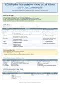
NURS 6023 Easy Study Guide | ECG Rhythm Interpretation + Intro to Lab Values
ECG Rhythm Interpretation + Intro to Lab Values
Easy-to-Learn Exam Study Guide
Focus: diseases/rhythms, findings, diagnostic tests, interpretation, and exam pearls
How to use this guide
Start with the quick comparison tables before reading the longer details.
For ECGs, identify rate, regularity, P waves, PR interval, and QRS width in that order every time.
For labs, do not just memorize “high” or “low.” Connect the lab to the likely body system and symptoms.
Use the “Exam Pearl” boxes as last-minute review before a quiz or diagnostic exam.
1. ECG Basics
Know the normal pathway and what each ECG part represents before memorizing rhythms.
Concept What it means Exam memory cue
Normal conduction “SA-AV-His-Branches-Purkinje”
pathway SA node -> AV node -> Bundle of His -> bundle branches -> Purkinje fibers
P wave P = “Push atria”
Atrial depolarization
Atrial contraction begins after this electrical signal
PR interval PR = “Pause before ventricles”
Atrial depolarization + AV junction delay
Normal: 0.12-0.20 sec or 3-5 small boxes
QRS complex Wide QRS usually means ventricular
Ventricular depolarization origin or bundle branch delay
Normal: 0.04-0.12 sec or 1-3 small boxes
T wave T = “Tired ventricles resting”
Ventricular repolarization
ECG paper 6-second strip: count R waves x 10
Small box = 0.04 sec
Large box = 0.20 sec
15 large boxes = 3 seconds
Normal pacemaker rates
SA node: 60-100 bpm. This is the dominant pacemaker.
AV node: 40-60 bpm. Backup pacemaker.
Ventricular cells: about 20-45 bpm. Slow backup; usually poor perfusion if relied on.
5-Step Rhythm Analysis: Use Every Time
Step Question to ask Normal answer
1. Rate How fast is it? 60-100 bpm for normal sinus rhythm
2. Regularity Are the R-R intervals evenly spaced? Regular
3. P waves Are P waves present, similar, and before each QRS? One normal P before every QRS
4. PR interval Is PR 0.12-0.20 sec? 3-5 small boxes
5. QRS duration Is QRS 0.04-0.12 sec? 1-3 small boxes; narrow = supraventricular conduction
2. Rhythm Recognition Cheat Sheet
High-yield “what makes it different” features.
Rhythm Key ECG findings Interpretation / why it happens Exam pearl
Normal Sinus Rhythm Impulse starts in SA node and conducts All 5 rhythm-analysis steps are
Rate 60-100 normally. normal.
Regular
Normal P before every QRS
PR 0.12-0.20
QRS 0.04-0.12
Study focus: findings, diagnostics, interpretation, exam pearls
, NURS 6023 Easy Study Guide | ECG Rhythm Interpretation + Intro to Lab Values
Rhythm Key ECG findings Interpretation / why it happens Exam pearl
Sinus Bradycardia SA node fires slower than normal but Only abnormality is the rate.
Rate < 60 conduction is normal.
Regular
Normal P/PR/QRS
Sinus Tachycardia SA node fires faster; often response to stress, Treat the cause, not just the
Rate > 100 fever, pain, hypovolemia, anxiety, hypoxia. rate.
Regular
Normal P/PR/QRS
PAC Ectopic atrial impulse conducts normally Atrial origin = usually narrow
Early beat through ventricles. QRS.
P wave shape different
QRS usually narrow
PVC Ventricular cell fires early; ventricular Ventricular origin = wide/bizarre
Early wide, bizarre QRS conduction is inefficient. QRS.
No preceding P for that beat
Atrial Fibrillation Chaotic atrial activity; AV node allows No P waves + irregularly
Irregularly irregular impulses through unpredictably. irregular = Afib. Think stroke
No organized P waves risk.
Narrow QRS usually
Atrial Flutter Re-entry circuit in right atrium; every Sawtooth = flutter.
Sawtooth flutter waves 2nd/3rd/4th impulse may conduct.
Atrial rate often 250-350
QRS usually narrow
PSVT Usually re-entry above ventricles, often Paroxysmal = starts/stops
Sudden fast regular rhythm around AV node. suddenly. Narrow QRS = above
P waves may disappear ventricles.
Narrow QRS
Ventricular Tachycardia Ventricular re-entry loop; may or may not Wide regular tachycardia =
Rate often fast have a pulse. treat as VT until proven
Regular otherwise.
No P waves
Wide QRS > 0.12
Ventricular Fibrillation Ventricular cells depolarize randomly; cardiac Deadly rhythm. Needs
Chaotic, no organized complexes output rapidly drops. immediate reversal.
No measurable rate/PR/QRS
AV Blocks: Compare the PR Pattern
Block ECG finding What is happening Memory cue
1st Degree AV Block PR interval > 0.20 sec; every P still has a QRS. Delayed conduction through AV “Long PR, but all get
node/Bundle of His. through.”
2nd Degree Type I / PR gets longer, longer, longer, then a QRS drops. Each impulse has more AV delay until “Longer, longer, drop.”
Wenckebach one fails.
2nd Degree Type II PR stays the same, but some P waves do not All-or-nothing conduction; often lower “Random dropped QRS
conduct to QRS. conduction system. with fixed PR.” More
concerning.
3rd Degree / Complete P waves and QRS complexes have no relationship; Atria and ventricles beat independently. “Divorced P and QRS.”
Heart Block ventricular escape rhythm may be wide/slow.
ECG “wide vs narrow” shortcut
Narrow QRS usually means the impulse came from above the ventricles and traveled normally through the ventricles.
Wide QRS means ventricular origin OR delayed ventricular conduction, such as bundle branch block.
PVC, VT, ventricular escape rhythms, and bundle branch blocks tend to have wide QRS complexes.
3. 12-Lead ECG: MI Location & Advanced Basics
Use the leads to locate the affected heart wall.
ECG finding Interpretation Exam pearl
ST elevation > 1 small box in 2 related Consistent with acute myocardial infarction in the matching heart Need 12-lead ECG, not just a rhythm strip.
leads region.
ST depression + T-wave inversion May indicate ischemia or non-ST elevation infarction pattern NSTEMI can have ST depression and T-
depending on clinical context. wave inversion.
Pathologic Q waves Can appear with infarction and may persist after fibrosis/scarring. Q waves may remain even when ST/T
normalize.
Tall QRS voltage Suggests left ventricular hypertrophy. LVH = thick muscle = bigger electrical
forces.
QRS > 0.12 with morphology change Bundle branch block pattern. BBB widens QRS because ventricular
conduction is delayed.
Study focus: findings, diagnostics, interpretation, exam pearls
ECG Rhythm Interpretation + Intro to Lab Values
Easy-to-Learn Exam Study Guide
Focus: diseases/rhythms, findings, diagnostic tests, interpretation, and exam pearls
How to use this guide
Start with the quick comparison tables before reading the longer details.
For ECGs, identify rate, regularity, P waves, PR interval, and QRS width in that order every time.
For labs, do not just memorize “high” or “low.” Connect the lab to the likely body system and symptoms.
Use the “Exam Pearl” boxes as last-minute review before a quiz or diagnostic exam.
1. ECG Basics
Know the normal pathway and what each ECG part represents before memorizing rhythms.
Concept What it means Exam memory cue
Normal conduction “SA-AV-His-Branches-Purkinje”
pathway SA node -> AV node -> Bundle of His -> bundle branches -> Purkinje fibers
P wave P = “Push atria”
Atrial depolarization
Atrial contraction begins after this electrical signal
PR interval PR = “Pause before ventricles”
Atrial depolarization + AV junction delay
Normal: 0.12-0.20 sec or 3-5 small boxes
QRS complex Wide QRS usually means ventricular
Ventricular depolarization origin or bundle branch delay
Normal: 0.04-0.12 sec or 1-3 small boxes
T wave T = “Tired ventricles resting”
Ventricular repolarization
ECG paper 6-second strip: count R waves x 10
Small box = 0.04 sec
Large box = 0.20 sec
15 large boxes = 3 seconds
Normal pacemaker rates
SA node: 60-100 bpm. This is the dominant pacemaker.
AV node: 40-60 bpm. Backup pacemaker.
Ventricular cells: about 20-45 bpm. Slow backup; usually poor perfusion if relied on.
5-Step Rhythm Analysis: Use Every Time
Step Question to ask Normal answer
1. Rate How fast is it? 60-100 bpm for normal sinus rhythm
2. Regularity Are the R-R intervals evenly spaced? Regular
3. P waves Are P waves present, similar, and before each QRS? One normal P before every QRS
4. PR interval Is PR 0.12-0.20 sec? 3-5 small boxes
5. QRS duration Is QRS 0.04-0.12 sec? 1-3 small boxes; narrow = supraventricular conduction
2. Rhythm Recognition Cheat Sheet
High-yield “what makes it different” features.
Rhythm Key ECG findings Interpretation / why it happens Exam pearl
Normal Sinus Rhythm Impulse starts in SA node and conducts All 5 rhythm-analysis steps are
Rate 60-100 normally. normal.
Regular
Normal P before every QRS
PR 0.12-0.20
QRS 0.04-0.12
Study focus: findings, diagnostics, interpretation, exam pearls
, NURS 6023 Easy Study Guide | ECG Rhythm Interpretation + Intro to Lab Values
Rhythm Key ECG findings Interpretation / why it happens Exam pearl
Sinus Bradycardia SA node fires slower than normal but Only abnormality is the rate.
Rate < 60 conduction is normal.
Regular
Normal P/PR/QRS
Sinus Tachycardia SA node fires faster; often response to stress, Treat the cause, not just the
Rate > 100 fever, pain, hypovolemia, anxiety, hypoxia. rate.
Regular
Normal P/PR/QRS
PAC Ectopic atrial impulse conducts normally Atrial origin = usually narrow
Early beat through ventricles. QRS.
P wave shape different
QRS usually narrow
PVC Ventricular cell fires early; ventricular Ventricular origin = wide/bizarre
Early wide, bizarre QRS conduction is inefficient. QRS.
No preceding P for that beat
Atrial Fibrillation Chaotic atrial activity; AV node allows No P waves + irregularly
Irregularly irregular impulses through unpredictably. irregular = Afib. Think stroke
No organized P waves risk.
Narrow QRS usually
Atrial Flutter Re-entry circuit in right atrium; every Sawtooth = flutter.
Sawtooth flutter waves 2nd/3rd/4th impulse may conduct.
Atrial rate often 250-350
QRS usually narrow
PSVT Usually re-entry above ventricles, often Paroxysmal = starts/stops
Sudden fast regular rhythm around AV node. suddenly. Narrow QRS = above
P waves may disappear ventricles.
Narrow QRS
Ventricular Tachycardia Ventricular re-entry loop; may or may not Wide regular tachycardia =
Rate often fast have a pulse. treat as VT until proven
Regular otherwise.
No P waves
Wide QRS > 0.12
Ventricular Fibrillation Ventricular cells depolarize randomly; cardiac Deadly rhythm. Needs
Chaotic, no organized complexes output rapidly drops. immediate reversal.
No measurable rate/PR/QRS
AV Blocks: Compare the PR Pattern
Block ECG finding What is happening Memory cue
1st Degree AV Block PR interval > 0.20 sec; every P still has a QRS. Delayed conduction through AV “Long PR, but all get
node/Bundle of His. through.”
2nd Degree Type I / PR gets longer, longer, longer, then a QRS drops. Each impulse has more AV delay until “Longer, longer, drop.”
Wenckebach one fails.
2nd Degree Type II PR stays the same, but some P waves do not All-or-nothing conduction; often lower “Random dropped QRS
conduct to QRS. conduction system. with fixed PR.” More
concerning.
3rd Degree / Complete P waves and QRS complexes have no relationship; Atria and ventricles beat independently. “Divorced P and QRS.”
Heart Block ventricular escape rhythm may be wide/slow.
ECG “wide vs narrow” shortcut
Narrow QRS usually means the impulse came from above the ventricles and traveled normally through the ventricles.
Wide QRS means ventricular origin OR delayed ventricular conduction, such as bundle branch block.
PVC, VT, ventricular escape rhythms, and bundle branch blocks tend to have wide QRS complexes.
3. 12-Lead ECG: MI Location & Advanced Basics
Use the leads to locate the affected heart wall.
ECG finding Interpretation Exam pearl
ST elevation > 1 small box in 2 related Consistent with acute myocardial infarction in the matching heart Need 12-lead ECG, not just a rhythm strip.
leads region.
ST depression + T-wave inversion May indicate ischemia or non-ST elevation infarction pattern NSTEMI can have ST depression and T-
depending on clinical context. wave inversion.
Pathologic Q waves Can appear with infarction and may persist after fibrosis/scarring. Q waves may remain even when ST/T
normalize.
Tall QRS voltage Suggests left ventricular hypertrophy. LVH = thick muscle = bigger electrical
forces.
QRS > 0.12 with morphology change Bundle branch block pattern. BBB widens QRS because ventricular
conduction is delayed.
Study focus: findings, diagnostics, interpretation, exam pearls


