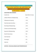
APEA PATHOPHYSIOLOGY ACTUAL EXAM REVIEW
2026 LATEST EDITION 180 QUESTIONS AND
CORRECT ANSWERS WITH RATIONALES
GUARANTEE A+ GRADE
EXAM STRUCTURE OVERVIEW
Content Area Approximate Coverage
Cellular & Molecular Pathophysiology ~15%
Inflammation & Immunity ~15%
Fluid, Electrolyte & Acid-Base ~10%
Hematologic Pathophysiology ~10%
Cardiovascular Pathophysiology ~15%
Respiratory Pathophysiology ~10%
Renal & Genitourinary ~8%
Gastrointestinal ~7%
Neurological ~5%
Endocrine ~5%
SECTION 1: CELLULAR & MOLECULAR PATHOPHYSIOLOGY
,Question 1
A 58-year-old patient with chronic gastroesophageal reflux disease undergoes
an endoscopy revealing that the squamous epithelium of the lower esophagus
has been replaced by columnar epithelium. This cellular adaptation is best
described as:
A. Hyperplasia
B. Metaplasia
C. Dysplasia
D. Anaplasia
Correct answer: B
Rationale: Metaplasia is a reversible change in which one differentiated cell
type is replaced by another. In Barrett's esophagus, the squamous epithelium
is replaced by columnar epithelium as an adaptive response to chronic acid
exposure. Hyperplasia is an increase in cell number, dysplasia involves
abnormal cell growth, and anaplasia refers to undifferentiated malignant cells.
Question 2
A 65-year-old patient with a history of smoking presents with a lung mass.
Biopsy reveals cells that are abnormal in size, shape, and organization. This
finding is most consistent with:
A. Hyperplasia
B. Metaplasia
C. Dysplasia
D. Anaplasia
Correct answer: C
Rationale: Dysplasia is characterized by abnormal changes in cell size, shape,
and organization. It is often a precancerous condition. Anaplasia refers to
undifferentiated cells seen in malignant tumors.
Question 3
,Which of the following best describes the mechanism of cellular injury in
ischemia?
A. Direct damage to cell membranes by free radicals
B. Lack of oxygen leading to ATP depletion and failure of sodium-potassium
pumps
C. Activation of the complement system
D. Accumulation of misfolded proteins in the endoplasmic reticulum
Correct answer: B
Rationale: Ischemia causes cellular injury primarily through oxygen
deprivation, leading to decreased ATP production. This results in failure of the
Na+/K+ ATPase pump, intracellular sodium accumulation, cellular swelling,
and ultimately cell death. Free radical damage and complement activation are
mechanisms of other types of injury.
Question 4
A patient with hereditary hemochromatosis has excessive iron deposition in
various organs. The type of cellular accumulation seen in this condition is:
A. Lipofuscin accumulation
B. Glycogen accumulation
C. Hemosiderin accumulation
D. Calcium accumulation
Correct answer: C
Rationale: Hemosiderin is an iron-containing pigment that accumulates in
tissues when there is excess iron. Hereditary hemochromatosis causes
widespread hemosiderin deposition in organs such as the liver, pancreas, and
heart.
Question 5
A 72-year-old patient with Alzheimer's disease has intracellular
accumulations of abnormal proteins. These accumulations are most likely:
A. Glycogen
B. Lipofuscin
, C. Neurofibrillary tangles
D. Mallory bodies
Correct answer: C
Rationale: Neurofibrillary tangles are intracellular accumulations of
hyperphosphorylated tau protein found in Alzheimer's disease. Lipofuscin is
"wear-and-tear" pigment, Mallory bodies are found in alcoholic liver disease,
and glycogen accumulation occurs in glycogen storage diseases.
Question 6
Which type of necrosis is most commonly associated with ischemic injury to
the heart?
A. Coagulative necrosis
B. Liquefactive necrosis
C. Caseous necrosis
D. Fat necrosis
Correct answer: A
Rationale: Coagulative necrosis is the characteristic pattern seen in ischemic
injury to solid organs, particularly the heart and kidneys. It preserves tissue
architecture for several days due to denaturation of proteins. Liquefactive
necrosis occurs in the brain, caseous necrosis in tuberculosis, and fat necrosis
in pancreatic injury.
Question 7
A 45-year-old patient with a prolonged fever develops a brain abscess. The
type of necrosis most likely seen in this lesion is:
A. Coagulative necrosis
B. Liquefactive necrosis
C. Caseous necrosis
D. Gangrenous necrosis
Correct answer: B
2026 LATEST EDITION 180 QUESTIONS AND
CORRECT ANSWERS WITH RATIONALES
GUARANTEE A+ GRADE
EXAM STRUCTURE OVERVIEW
Content Area Approximate Coverage
Cellular & Molecular Pathophysiology ~15%
Inflammation & Immunity ~15%
Fluid, Electrolyte & Acid-Base ~10%
Hematologic Pathophysiology ~10%
Cardiovascular Pathophysiology ~15%
Respiratory Pathophysiology ~10%
Renal & Genitourinary ~8%
Gastrointestinal ~7%
Neurological ~5%
Endocrine ~5%
SECTION 1: CELLULAR & MOLECULAR PATHOPHYSIOLOGY
,Question 1
A 58-year-old patient with chronic gastroesophageal reflux disease undergoes
an endoscopy revealing that the squamous epithelium of the lower esophagus
has been replaced by columnar epithelium. This cellular adaptation is best
described as:
A. Hyperplasia
B. Metaplasia
C. Dysplasia
D. Anaplasia
Correct answer: B
Rationale: Metaplasia is a reversible change in which one differentiated cell
type is replaced by another. In Barrett's esophagus, the squamous epithelium
is replaced by columnar epithelium as an adaptive response to chronic acid
exposure. Hyperplasia is an increase in cell number, dysplasia involves
abnormal cell growth, and anaplasia refers to undifferentiated malignant cells.
Question 2
A 65-year-old patient with a history of smoking presents with a lung mass.
Biopsy reveals cells that are abnormal in size, shape, and organization. This
finding is most consistent with:
A. Hyperplasia
B. Metaplasia
C. Dysplasia
D. Anaplasia
Correct answer: C
Rationale: Dysplasia is characterized by abnormal changes in cell size, shape,
and organization. It is often a precancerous condition. Anaplasia refers to
undifferentiated cells seen in malignant tumors.
Question 3
,Which of the following best describes the mechanism of cellular injury in
ischemia?
A. Direct damage to cell membranes by free radicals
B. Lack of oxygen leading to ATP depletion and failure of sodium-potassium
pumps
C. Activation of the complement system
D. Accumulation of misfolded proteins in the endoplasmic reticulum
Correct answer: B
Rationale: Ischemia causes cellular injury primarily through oxygen
deprivation, leading to decreased ATP production. This results in failure of the
Na+/K+ ATPase pump, intracellular sodium accumulation, cellular swelling,
and ultimately cell death. Free radical damage and complement activation are
mechanisms of other types of injury.
Question 4
A patient with hereditary hemochromatosis has excessive iron deposition in
various organs. The type of cellular accumulation seen in this condition is:
A. Lipofuscin accumulation
B. Glycogen accumulation
C. Hemosiderin accumulation
D. Calcium accumulation
Correct answer: C
Rationale: Hemosiderin is an iron-containing pigment that accumulates in
tissues when there is excess iron. Hereditary hemochromatosis causes
widespread hemosiderin deposition in organs such as the liver, pancreas, and
heart.
Question 5
A 72-year-old patient with Alzheimer's disease has intracellular
accumulations of abnormal proteins. These accumulations are most likely:
A. Glycogen
B. Lipofuscin
, C. Neurofibrillary tangles
D. Mallory bodies
Correct answer: C
Rationale: Neurofibrillary tangles are intracellular accumulations of
hyperphosphorylated tau protein found in Alzheimer's disease. Lipofuscin is
"wear-and-tear" pigment, Mallory bodies are found in alcoholic liver disease,
and glycogen accumulation occurs in glycogen storage diseases.
Question 6
Which type of necrosis is most commonly associated with ischemic injury to
the heart?
A. Coagulative necrosis
B. Liquefactive necrosis
C. Caseous necrosis
D. Fat necrosis
Correct answer: A
Rationale: Coagulative necrosis is the characteristic pattern seen in ischemic
injury to solid organs, particularly the heart and kidneys. It preserves tissue
architecture for several days due to denaturation of proteins. Liquefactive
necrosis occurs in the brain, caseous necrosis in tuberculosis, and fat necrosis
in pancreatic injury.
Question 7
A 45-year-old patient with a prolonged fever develops a brain abscess. The
type of necrosis most likely seen in this lesion is:
A. Coagulative necrosis
B. Liquefactive necrosis
C. Caseous necrosis
D. Gangrenous necrosis
Correct answer: B