MSCI 510 Test 2 Study Guide with Complete Solution | Latest Update 2026–
2027
The Thorax
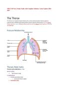
The thorax is an irregularly shaped cylinder with a narrow opening (superior thoracic aperture)
superiorly and a relatively large opening (inferior thoracic aperture) inferiorly (Fig. 3.1). The superior
thoracic aperture is open, allowing continuity with the neck; the inferior thoracic aperture is closed by
the diaphragm.
Pressure Relationship:
Thoracic Body Cavity
Thoracic cavity subdivisions: a. Two
pleural cavities
• Each houses a lung
b. Mediastinum
• Contains pericardial cavity
– Surrounds thoracic organs
• Pericardial cavity
, – Encloses heart
Mediastinum:
The mediastinum is a thick, flexible soft tissue partition oriented longitudinally in a median sagittal
position. It contains the heart, esophagus, trachea, major nerves, and major systemic blood vessels.
Pleural Cavities (Right and Left):
The pleural cavities are completely separated from each other by the mediastinum. Therefore, abnormal
events in one pleural cavity do not necessarily affect the other cavity. This also means that the
mediastinum can be entered surgically without opening the pleural cavities.
Another important feature of the pleural cavities is that they extend above the level of rib I. The apex of
each lung extends into the root of the neck. Consequently, abnormal events in the root of the neck can
involve the adjacent pleura and lung, and events in the adjacent pleura and lung can involve the root of
the neck.
Functions of the Thorax:
1. Breathing
2. Protection of vital organs
3. Conduit
Breathing: the thorax not only contains the lungs but also provides the machinery necessary—the
diaphragm, thoracic wall, and the ribs—for effectively moving air into and out of the lungs.
Up and down movements of the diaphragm and changes in the lateral and anterior dimensions of the
thoracic wall, caused by movements of the ribs, alter the volume of the thoracic cavity and are key
elements in breathing (Inhalation and Exhalation).
Protection of Vital Organs: houses and protects the heart, lungs, and great vessels. Because of the
domed shape of the diaphragm, the thoracic wall also offers protection to the liver lies under the right
dome of the diaphragm, and the stomach and spleen lie under the left. The posterior aspects of the
superior poles of the kidneys lie on the diaphragm and are anterior to rib XII, on the right, and to ribs XI
and XII, on the left.
Conduit: the mediastinum acts as a conduit for structures that pass completely through the thorax.
The esophagus, vagus nerves, thoracic duct pass the phrenic nerves, the trachea, thoracic aorta, and
superior vena cava course within the mediastinum.
Thoracic Wall Boundaries:
, Consists of Skeletal and Muscular elements
• Posteriorly, is made up ofo Twelve thoracic
vertebrae and their intervening intervertebral
discs
• Laterally, is made up ofo Ribs (twelve on
each side) o Three Layers of Flat Muscles,
which span the intercostal spaces between
adjacent ribs, move the ribs and provide
support for the intercostal spaces
• Anteriorly, is made up of theo Sternum
which consists of:
Manubrium
Body of Sternum
Xiphoid Process
Superior Thoracic Aperture
The first ribs slope inferiorly from their posterior
articulation with vertebra TI to their anterior
attachment to the manubrium. Consequently,
the plane of the superior thoracic aperture is at
an oblique angle, facing somewhat anteriorly.
The superior margin of the manubrium is in
approximately the same horizontal plane as the
intervertebral disc between vertebrae TII and
TIII.
Inferior Thoracic Aperture
The inferior thoracic aperture is large and expandable
and is closed by the diaphragm, and structures passing
between the abdomen and thorax pierce or pass posteriorly
to the diaphragm.
Four Main Apertures:
1. Esophageal
2. Thoracic Duct and
3. Aortic Duct TOGETHER
4. Inferior Vena Cava
Skeletal Elements of the Inferior Thoracic Aperture:
• The body of vertebra TXII posteriorly
• Rib XII and the distal end of rib XI posterolaterally
• Distal cartilaginous ends of ribs VII to X, which unite to form the costal margin anterolaterally
• Xiphoid process anteriorly.
2027
The Thorax
The thorax is an irregularly shaped cylinder with a narrow opening (superior thoracic aperture)
superiorly and a relatively large opening (inferior thoracic aperture) inferiorly (Fig. 3.1). The superior
thoracic aperture is open, allowing continuity with the neck; the inferior thoracic aperture is closed by
the diaphragm.
Pressure Relationship:
Thoracic Body Cavity
Thoracic cavity subdivisions: a. Two
pleural cavities
• Each houses a lung
b. Mediastinum
• Contains pericardial cavity
– Surrounds thoracic organs
• Pericardial cavity
, – Encloses heart
Mediastinum:
The mediastinum is a thick, flexible soft tissue partition oriented longitudinally in a median sagittal
position. It contains the heart, esophagus, trachea, major nerves, and major systemic blood vessels.
Pleural Cavities (Right and Left):
The pleural cavities are completely separated from each other by the mediastinum. Therefore, abnormal
events in one pleural cavity do not necessarily affect the other cavity. This also means that the
mediastinum can be entered surgically without opening the pleural cavities.
Another important feature of the pleural cavities is that they extend above the level of rib I. The apex of
each lung extends into the root of the neck. Consequently, abnormal events in the root of the neck can
involve the adjacent pleura and lung, and events in the adjacent pleura and lung can involve the root of
the neck.
Functions of the Thorax:
1. Breathing
2. Protection of vital organs
3. Conduit
Breathing: the thorax not only contains the lungs but also provides the machinery necessary—the
diaphragm, thoracic wall, and the ribs—for effectively moving air into and out of the lungs.
Up and down movements of the diaphragm and changes in the lateral and anterior dimensions of the
thoracic wall, caused by movements of the ribs, alter the volume of the thoracic cavity and are key
elements in breathing (Inhalation and Exhalation).
Protection of Vital Organs: houses and protects the heart, lungs, and great vessels. Because of the
domed shape of the diaphragm, the thoracic wall also offers protection to the liver lies under the right
dome of the diaphragm, and the stomach and spleen lie under the left. The posterior aspects of the
superior poles of the kidneys lie on the diaphragm and are anterior to rib XII, on the right, and to ribs XI
and XII, on the left.
Conduit: the mediastinum acts as a conduit for structures that pass completely through the thorax.
The esophagus, vagus nerves, thoracic duct pass the phrenic nerves, the trachea, thoracic aorta, and
superior vena cava course within the mediastinum.
Thoracic Wall Boundaries:
, Consists of Skeletal and Muscular elements
• Posteriorly, is made up ofo Twelve thoracic
vertebrae and their intervening intervertebral
discs
• Laterally, is made up ofo Ribs (twelve on
each side) o Three Layers of Flat Muscles,
which span the intercostal spaces between
adjacent ribs, move the ribs and provide
support for the intercostal spaces
• Anteriorly, is made up of theo Sternum
which consists of:
Manubrium
Body of Sternum
Xiphoid Process
Superior Thoracic Aperture
The first ribs slope inferiorly from their posterior
articulation with vertebra TI to their anterior
attachment to the manubrium. Consequently,
the plane of the superior thoracic aperture is at
an oblique angle, facing somewhat anteriorly.
The superior margin of the manubrium is in
approximately the same horizontal plane as the
intervertebral disc between vertebrae TII and
TIII.
Inferior Thoracic Aperture
The inferior thoracic aperture is large and expandable
and is closed by the diaphragm, and structures passing
between the abdomen and thorax pierce or pass posteriorly
to the diaphragm.
Four Main Apertures:
1. Esophageal
2. Thoracic Duct and
3. Aortic Duct TOGETHER
4. Inferior Vena Cava
Skeletal Elements of the Inferior Thoracic Aperture:
• The body of vertebra TXII posteriorly
• Rib XII and the distal end of rib XI posterolaterally
• Distal cartilaginous ends of ribs VII to X, which unite to form the costal margin anterolaterally
• Xiphoid process anteriorly.