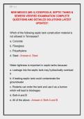
ROBBINS-INSPIRED PATHOLOGY EXAM
PREP
Advanced Clinical MCQs + Integrated Rationales + Higher-
Order Pathophysiology
Designed for learners seeking deeper clinical understanding beyond memorization-
heavy review materials
1. A 24-year-old woman presents with progressive periorbital
edema and frothy urine two weeks after an upper
respiratory infection. Laboratory studies reveal severe
proteinuria, hypoalbuminemia, and hyperlipidemia. Renal
biopsy demonstrates diffuse effacement of podocyte foot
processes without immune complex deposition.
Which pathophysiologic alteration most directly
predisposes this patient to venous thrombosis?
A. Increased hepatic fibrinogen degradation
B. Urinary loss of antithrombin III
,C. Complement-mediated endothelial destruction
D. Reduced platelet adhesion factor synthesis
E. Impaired hepatic coagulation factor production
Correct Answer: B. Urinary loss of antithrombin III
Key Diagnostic Clue
The combination of massive proteinuria, edema, and
selective albumin loss indicates nephrotic syndrome, most
consistent with minimal change disease.
Mechanistic Interpretation
Nephrotic syndromes cause urinary loss of low-molecular-
weight anticoagulant proteins, especially antithrombin III.
This shifts hemostatic balance toward hypercoagulability.
Why the Disease Behaves This Way
The liver compensates for urinary protein loss by increasing
synthesis of coagulation factors, worsening thrombotic
tendency despite low serum albumin.
Why Other Choices Fail
• A. Fibrinogen degradation would reduce clotting, not
enhance it.
• C. Endothelial destruction is not the primary mechanism of
thrombosis here.
• D. Platelet adhesion is generally preserved.
,• E. Hepatic coagulation factor synthesis is increased, not
impaired.
Exam Trap
Students often associate edema with nephritic syndromes
and overlook the hypercoagulable complications unique to
nephrotic disease.
Clinical Correlation
Renal vein thrombosis is a classic complication of severe
nephrotic syndrome.
Memory Anchor
“Nephrotic urine loses anticoagulants before clotting
factors.”
2. A 67-year-old man with a 50-pack-year smoking history
presents with persistent cough, hemoptysis, and weight
loss. Imaging reveals a centrally located hilar mass.
Laboratory studies show hypercalcemia with suppressed
parathyroid hormone levels.
Which mechanism most directly explains this patient’s
metabolic abnormality?
A. Osteolytic cytokine release from bone metastases
B. Ectopic secretion of parathyroid hormone–related peptide
C. Vitamin D activation by tumor macrophages
, D. Increased calcitonin receptor resistance
E. Tumor-induced adrenal insufficiency
Correct Answer: B. Ectopic secretion of parathyroid
hormone–related peptide
Key Diagnostic Clue
A central hilar lung mass with hypercalcemia strongly
suggests squamous cell carcinoma of the lung.
Mechanistic Interpretation
Squamous cell carcinomas commonly produce PTHrP, which
mimics parathyroid hormone activity and increases serum
calcium.
Why Correct Answer Wins
PTHrP enhances osteoclastic bone resorption and renal
calcium reabsorption while suppressing endogenous PTH.
Why Other Choices Fail
• A. Osteolytic metastases can cause hypercalcemia but are
less characteristic here.
• C. Vitamin D activation is classically seen in granulomatous
disease.
• D. Calcitonin resistance is unrelated.
• E. Adrenal insufficiency does not produce marked
hypercalcemia via this mechanism.
Exam Pearl