

Lab Kit-Mitosis and Meiosis: Chromosome Simulation
Pre-Lab Questions:
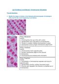
1. Identify the stages of mitosis in the following photomicrograph of histological
slides and list the events happening in each phase:
Stages of Mitosis
Phase: Metaphase
Events:
1. Chromosomes line up at the cell's center.
2. Spindle fibers form and attach to chromosomes.
3. Tension is created, holding chromosomes in place.
4. Checkpoints ensure proper chromosome attachment.
5. This sets the stage for chromosome separation in
Phase: Telophase
Events:
1. Chromosomes reach opposite poles.
2. Nuclear membranes begin to form around
chromosomes.
3. Chromosomes de-condense into chromatin.
4. Spindle fibers disappear.
Phase: Anaphase
Events:
1. Chromatids or chromosomes separate and move to
opposite poles.
2. Spindle fibers shorten, pulling chromatids apart.
3. Ensures each daughter cell receives a complete set of
chromosomes.
, 1
Images of Mitosis Models:
Photo 1-Interphase
Insert the photo from Activity 1, step 3d. The photo should be of the model (including the
label) of Interphase (Before Mitosis).
, 2
Photo 2- Prophase
Insert the photo from Activity 1, step 4e. The photo should be of the model (including the
label) of Prophase.
Pre-Lab Questions:
1. Identify the stages of mitosis in the following photomicrograph of histological
slides and list the events happening in each phase:
Stages of Mitosis
Phase: Metaphase
Events:
1. Chromosomes line up at the cell's center.
2. Spindle fibers form and attach to chromosomes.
3. Tension is created, holding chromosomes in place.
4. Checkpoints ensure proper chromosome attachment.
5. This sets the stage for chromosome separation in
Phase: Telophase
Events:
1. Chromosomes reach opposite poles.
2. Nuclear membranes begin to form around
chromosomes.
3. Chromosomes de-condense into chromatin.
4. Spindle fibers disappear.
Phase: Anaphase
Events:
1. Chromatids or chromosomes separate and move to
opposite poles.
2. Spindle fibers shorten, pulling chromatids apart.
3. Ensures each daughter cell receives a complete set of
chromosomes.
, 1
Images of Mitosis Models:
Photo 1-Interphase
Insert the photo from Activity 1, step 3d. The photo should be of the model (including the
label) of Interphase (Before Mitosis).
, 2
Photo 2- Prophase
Insert the photo from Activity 1, step 4e. The photo should be of the model (including the
label) of Prophase.


