

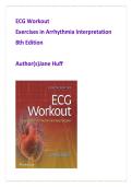
ECG Workout
Exercises in Arrhythmia Interpretation
8th Edition
Author(s)Jane Huff
,TEST BANK
Table of Contents — ECG Workout: Exercises in Arrhythmia Interpretation, 8th Edition
1. Anatomy and Physiology of the Heart
2. Electrophysiology
3. Waveforms, Complexes, Intervals, and Segments
4. Cardiac Monitors
5. Analyzing a Rhythm Strip
6. Sinus Rhythms
7. Atrial Rhythms
8. Junctional Rhythms and AV Blocks
9. Ventricular Rhythms and Bundle-Branch Block
10. Pacemakers
11. Posttest
Question 1
An ECG shows a regular rhythm at 72 bpm with upright P waves
preceding each QRS complex. The nurse recalls that proper
valve function ensures unidirectional blood flow. Which valve
prevents backflow from the left ventricle into the left atrium
during ventricular contraction?
A. Tricuspid valve
B. Pulmonic valve
C. Mitral valve
D. Aortic valve
,Correct Answer: C
Rationale:
The mitral valve closes during ventricular systole to prevent
backflow into the left atrium. The ECG reflects normal sinus
rhythm, indicating coordinated atrial and ventricular
contraction.
• A: Tricuspid valve is on the right side
• B: Pulmonic valve prevents backflow into right ventricle
• D: Aortic valve prevents backflow into left ventricle
Source: ECG Workout: Exercises in Arrhythmia Interpretation,
8th Edition – Chapter 1
Question 2
An ECG rhythm shows consistent P-QRS-T cycles at 80 bpm. The
nurse understands that blood leaving the right ventricle flows
into which structure next?
A. Aorta
B. Pulmonary artery
C. Left atrium
D. Superior vena cava
Correct Answer: B
Rationale:
Blood flows from the right ventricle through the pulmonic valve
, into the pulmonary artery toward the lungs. The ECG indicates
effective conduction supporting normal flow.
• A: Aorta carries blood from left ventricle
• C: Left atrium receives oxygenated blood
• D: Superior vena cava returns systemic blood
Source: ECG Workout: Exercises in Arrhythmia Interpretation,
8th Edition – Chapter 1
Question 3
A patient’s ECG demonstrates a normal sinus rhythm at 68
bpm. Which structure is responsible for delivering oxygenated
blood from the lungs to the heart?
A. Pulmonary artery
B. Pulmonary vein
C. Inferior vena cava
D. Coronary sinus
Correct Answer: B
Rationale:
Pulmonary veins carry oxygenated blood from the lungs to the
left atrium. A normal ECG suggests adequate cardiac perfusion
and circulation.
• A: Carries deoxygenated blood to lungs
• C: Returns systemic venous blood
Exercises in Arrhythmia Interpretation
8th Edition
Author(s)Jane Huff
,TEST BANK
Table of Contents — ECG Workout: Exercises in Arrhythmia Interpretation, 8th Edition
1. Anatomy and Physiology of the Heart
2. Electrophysiology
3. Waveforms, Complexes, Intervals, and Segments
4. Cardiac Monitors
5. Analyzing a Rhythm Strip
6. Sinus Rhythms
7. Atrial Rhythms
8. Junctional Rhythms and AV Blocks
9. Ventricular Rhythms and Bundle-Branch Block
10. Pacemakers
11. Posttest
Question 1
An ECG shows a regular rhythm at 72 bpm with upright P waves
preceding each QRS complex. The nurse recalls that proper
valve function ensures unidirectional blood flow. Which valve
prevents backflow from the left ventricle into the left atrium
during ventricular contraction?
A. Tricuspid valve
B. Pulmonic valve
C. Mitral valve
D. Aortic valve
,Correct Answer: C
Rationale:
The mitral valve closes during ventricular systole to prevent
backflow into the left atrium. The ECG reflects normal sinus
rhythm, indicating coordinated atrial and ventricular
contraction.
• A: Tricuspid valve is on the right side
• B: Pulmonic valve prevents backflow into right ventricle
• D: Aortic valve prevents backflow into left ventricle
Source: ECG Workout: Exercises in Arrhythmia Interpretation,
8th Edition – Chapter 1
Question 2
An ECG rhythm shows consistent P-QRS-T cycles at 80 bpm. The
nurse understands that blood leaving the right ventricle flows
into which structure next?
A. Aorta
B. Pulmonary artery
C. Left atrium
D. Superior vena cava
Correct Answer: B
Rationale:
Blood flows from the right ventricle through the pulmonic valve
, into the pulmonary artery toward the lungs. The ECG indicates
effective conduction supporting normal flow.
• A: Aorta carries blood from left ventricle
• C: Left atrium receives oxygenated blood
• D: Superior vena cava returns systemic blood
Source: ECG Workout: Exercises in Arrhythmia Interpretation,
8th Edition – Chapter 1
Question 3
A patient’s ECG demonstrates a normal sinus rhythm at 68
bpm. Which structure is responsible for delivering oxygenated
blood from the lungs to the heart?
A. Pulmonary artery
B. Pulmonary vein
C. Inferior vena cava
D. Coronary sinus
Correct Answer: B
Rationale:
Pulmonary veins carry oxygenated blood from the lungs to the
left atrium. A normal ECG suggests adequate cardiac perfusion
and circulation.
• A: Carries deoxygenated blood to lungs
• C: Returns systemic venous blood


