Foot and Ankle
Learning Objectives
1. Identify the anatomical joints of the hindfoot, midfoot and forefoot.
2. Apply basic biomechanical principles to a foot and ankle case at an
efficiency of ~80% or higher.
3. Demonstrate basic understanding of supination and pronation twist.
4. Demonstrate basic understanding of pronation, supination, DF, and PF
motions of the foot and ankle.
Ankle and Foot Function
Stability:
Provide a stable base of support for the body in a variety of WB postures without
excessive muscle activity and energy expenditure
Act as a rigid lever for effective push-off during gait
Mobility:
Dampening rotations imposed by the proximal joints of lower limb
Being flexible to absorb shock as foot hits the ground
Permit the foot to conform to changing and varied terrain
Osteokinematics and Planes of Motion
Dorsiflexion & plantarflexion
• Sagittal plane, coronal axis
• Includes toe flexion and extension
Inversion & eversion
• Frontal plane, AP axis
Abduction & adduction
• Transverse plane, vertical axis
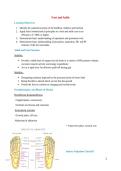
Surface Palpation Checkoff
1
, Navicular—Medial Column (Ray 1) Posterior Tibialis
Sustentaculum Tali
Calcaneonavicular ligament
Cuboid—Lateral Column (Ray 5) Peroneus Longus vs. Brevis Attachment
Tubercle of 5th metatarsal
Anterior Talofibular ligament
Talus Dorsalis Pedis Pulse
Cuneiforms (3)
Metatarsal
Functional Rays
A ray is a functional unit formed by a metatarsal and its associated cuneiform (1st– 3rd). The 4th
and 5th rays are just the metatarsal.
In biomechanics and movement science, the human foot has five rays, which are used to describe
the functional alignment and movement patterns of the foot. Each ray consists of a metatarsal
bone and its associated phalanges (toe bones). Here's a breakdown of the five rays:
First ray:
Comprised of the first metatarsal and the bones of the big toe (hallux).
This ray plays a crucial role in weight-bearing and propulsion during walking and
running.
Second ray:
Includes the second metatarsal and its associated phalanges.
Acts as the central stabilizer for the foot, supporting balance and distributing forces
during gait.
Third ray:
2
, Formed by the third metatarsal and its phalanges.
Functions in load distribution and stability.
Fourth ray:
Consists of the fourth metatarsal and its
phalanges.
Contributes to the flexibility and motion of
the lateral aspect of the foot.
Fifth ray:
Made up of the fifth metatarsal and the
bones of the small toe.
Provides balance and assists in lateral foot
movement, especially during activities that
involve side-to-side motion.
These rays are important in understanding how the
foot adapts to different movements and loads,
particularly in tasks like walking, running, and
maintaining balance. From a clinical perspective,
they are also important when considering foot
deformities, biomechanics of gait, and the effects of
weight distribution across the foot.
Joints Sections of the Foot
Foot/ankle complex
1. Distal tibiofibular joint
The distal tibiofibular joint is a syndesmosis, or fibrous union, between the concave
facet of the tibia and the convex facet of the fibula. The distal tibia and fibula do not
actually come into contact with each other but are separated by fibroadipose tissue.
2. Talocrural (Tibiotalar) joint
3. Subtalar joint
The subtalar joint articulating surfaces are highly variable, but the posterior articulation is
consistently the largest of the three articulations found between the talus and calcaneus.
The posterior articulation is formed by a concave facet on the undersurface of the body
of the talus and a convex facet on the body of the calcaneus. The smaller anterior and
middle talocalcaneal articulations are formed by two convex facets on the inferior body
and neck of the talus and two concave facets on the calcaneus.
3
Learning Objectives
1. Identify the anatomical joints of the hindfoot, midfoot and forefoot.
2. Apply basic biomechanical principles to a foot and ankle case at an
efficiency of ~80% or higher.
3. Demonstrate basic understanding of supination and pronation twist.
4. Demonstrate basic understanding of pronation, supination, DF, and PF
motions of the foot and ankle.
Ankle and Foot Function
Stability:
Provide a stable base of support for the body in a variety of WB postures without
excessive muscle activity and energy expenditure
Act as a rigid lever for effective push-off during gait
Mobility:
Dampening rotations imposed by the proximal joints of lower limb
Being flexible to absorb shock as foot hits the ground
Permit the foot to conform to changing and varied terrain
Osteokinematics and Planes of Motion
Dorsiflexion & plantarflexion
• Sagittal plane, coronal axis
• Includes toe flexion and extension
Inversion & eversion
• Frontal plane, AP axis
Abduction & adduction
• Transverse plane, vertical axis
Surface Palpation Checkoff
1
, Navicular—Medial Column (Ray 1) Posterior Tibialis
Sustentaculum Tali
Calcaneonavicular ligament
Cuboid—Lateral Column (Ray 5) Peroneus Longus vs. Brevis Attachment
Tubercle of 5th metatarsal
Anterior Talofibular ligament
Talus Dorsalis Pedis Pulse
Cuneiforms (3)
Metatarsal
Functional Rays
A ray is a functional unit formed by a metatarsal and its associated cuneiform (1st– 3rd). The 4th
and 5th rays are just the metatarsal.
In biomechanics and movement science, the human foot has five rays, which are used to describe
the functional alignment and movement patterns of the foot. Each ray consists of a metatarsal
bone and its associated phalanges (toe bones). Here's a breakdown of the five rays:
First ray:
Comprised of the first metatarsal and the bones of the big toe (hallux).
This ray plays a crucial role in weight-bearing and propulsion during walking and
running.
Second ray:
Includes the second metatarsal and its associated phalanges.
Acts as the central stabilizer for the foot, supporting balance and distributing forces
during gait.
Third ray:
2
, Formed by the third metatarsal and its phalanges.
Functions in load distribution and stability.
Fourth ray:
Consists of the fourth metatarsal and its
phalanges.
Contributes to the flexibility and motion of
the lateral aspect of the foot.
Fifth ray:
Made up of the fifth metatarsal and the
bones of the small toe.
Provides balance and assists in lateral foot
movement, especially during activities that
involve side-to-side motion.
These rays are important in understanding how the
foot adapts to different movements and loads,
particularly in tasks like walking, running, and
maintaining balance. From a clinical perspective,
they are also important when considering foot
deformities, biomechanics of gait, and the effects of
weight distribution across the foot.
Joints Sections of the Foot
Foot/ankle complex
1. Distal tibiofibular joint
The distal tibiofibular joint is a syndesmosis, or fibrous union, between the concave
facet of the tibia and the convex facet of the fibula. The distal tibia and fibula do not
actually come into contact with each other but are separated by fibroadipose tissue.
2. Talocrural (Tibiotalar) joint
3. Subtalar joint
The subtalar joint articulating surfaces are highly variable, but the posterior articulation is
consistently the largest of the three articulations found between the talus and calcaneus.
The posterior articulation is formed by a concave facet on the undersurface of the body
of the talus and a convex facet on the body of the calcaneus. The smaller anterior and
middle talocalcaneal articulations are formed by two convex facets on the inferior body
and neck of the talus and two concave facets on the calcaneus.
3