SPI EXAM REVIEW IMAGES EXAM QUESTIONS &
DETAILED CORRECT ANSWERS 100%
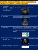
1. Decreased
The hyperechoic region (arrow) in this image results from:
attenua- tion
through a fluid
filled structure
2. Reflection What shadow depicted in this image of a renal stone is primarily a
result of the following sound tissue interaction:
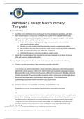
3. Scattering In the following illustration, if the propagation speed is unchanged at th
interface, what most correctly describes the sound tissue interaction that
will take place when the ultrasound wave strikes the depicted
interface?
4. Increased The dark area indicated by the arrow in this image is primarily due to
Attenua- tion
5. a lower frequency Picture A has than B. Each represents a signal versus tim
,SPI EXAM REVIEW IMAGES EXAM QUESTIONS &
DETAILED CORRECT ANSWERS 100%
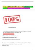
6. refraction
,SPI EXAM REVIEW IMAGES EXAM QUESTIONS &
DETAILED CORRECT ANSWERS 100%
Which of the following sound interactions produced the shadow indicated
by the arrows on this image?
7. acoustic What term describes the hyperechoic region (arrow) beneath this
enhance- ment complex structure?
8. Phased array What type of probe was used to take this image?
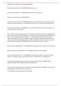
9. Increased line densi- The image on the left demonstrates poor lateral resolution compar
to the
ty image on the right. How were the system controls adjusted to
optimize the image on the right?
10. 3 In the image below, how many transmit focal zones are being used?
,SPI EXAM REVIEW IMAGES EXAM QUESTIONS &
DETAILED CORRECT ANSWERS 100%
DETAILED CORRECT ANSWERS 100%
1. Decreased
The hyperechoic region (arrow) in this image results from:
attenua- tion
through a fluid
filled structure
2. Reflection What shadow depicted in this image of a renal stone is primarily a
result of the following sound tissue interaction:
3. Scattering In the following illustration, if the propagation speed is unchanged at th
interface, what most correctly describes the sound tissue interaction that
will take place when the ultrasound wave strikes the depicted
interface?
4. Increased The dark area indicated by the arrow in this image is primarily due to
Attenua- tion
5. a lower frequency Picture A has than B. Each represents a signal versus tim
,SPI EXAM REVIEW IMAGES EXAM QUESTIONS &
DETAILED CORRECT ANSWERS 100%
6. refraction
,SPI EXAM REVIEW IMAGES EXAM QUESTIONS &
DETAILED CORRECT ANSWERS 100%
Which of the following sound interactions produced the shadow indicated
by the arrows on this image?
7. acoustic What term describes the hyperechoic region (arrow) beneath this
enhance- ment complex structure?
8. Phased array What type of probe was used to take this image?
9. Increased line densi- The image on the left demonstrates poor lateral resolution compar
to the
ty image on the right. How were the system controls adjusted to
optimize the image on the right?
10. 3 In the image below, how many transmit focal zones are being used?
,SPI EXAM REVIEW IMAGES EXAM QUESTIONS &
DETAILED CORRECT ANSWERS 100%