Week 7 Part 2 Study Guide
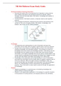
Renal Regulation of Acid-Base Balance
1. Source of Acidic By-Products:
- Cellular metabolism continuously produces acids that can disturb blood pH:
- Carbon dioxide from aerobic metabolism enters the bloodstream
- Hydrogen ions from glycolysis also enter the blood.
- Without buffering and excretion, these acids would lower blood pH dangerously.
2. Carbonic Anhydrase reaction:
- Inside red blood cells and renal tubular cells, enzyme carbonic anhydrase catalyzes a
reversible reaction.
- Key Points:
- Carbon dioxide and water combine to form carbonic acid which quickly dissociates into
hydrogen ions and bicarbonate
- Reverse reaction also occurs when buffering excess acid.
3. Kidney Handling of Hydrogen and Bicarbonate:
a. Bicarbonate Reabsorption:
- Bicarbonate is reabsorbed from filtrate back into the bloodstream.
- Increases plasma bicarbonate concentration- sometimes referred to as base excess.
- Extra bicarbonate acts as a buffer, neutralizing free hydrogen ions.
- Keeps blood pH stable around 7.35-7.45
b. Hydrogen Ion Secretion:
- Hydrogen ions are secreted from tubular cells into the filtrate.
- Thes hydrogen ions are then excreted in urine, effectively removing acid from the body.
Micturition Reflex (Urination reflex)
1. Two Basic Steps of Micturition:
- Bladder Filling:
- Bladder progressively fills with urine.
- Urine accumulates; bladder wall tension gradually increases.
- Tension rises above a threshold, activates stretch receptors in the bladder wall
- Reflex Emptying:
- Once threshold is reached, spinal cord reflex is triggered.
- Reflex leads to bladder contraction and emptying if conditions allow
2. Autonomic Control of the Bladder:
a. Sympathetic Nervous System
- Function: promotes urine retention
- Actions: relaxes detrusor muscle and contracts internal urethral sphincter
- Result: urine stays stored in the bladder
b. Parasympathetic Nervous System:
, - Function: promotes urination
- Actions: contracts detrusor muscle and relaxes internal urethral sphincter
- Result: urine flows out of the bladder into the urethra
3. Somatic Control:
- External urethral sphincter:
- Type: skeletal muscle
- Nerve supply: pudendal nerve
- Actions:
- Normally kept contracted to prevent urination
- Voluntarily relaxed when deciding to urinate
- Deactivation of sympathetic neurons
- Activation of parasympathetic neurons
4. Steps of Micturition reflex:
- 1. Bladder filling stretches the bladder wall
- 2. Stretch receptors in the bladder wall and posterior urethra send signals through afferent
fibers to the sacral spinal cord.
- 3. Reflex impulses are sent back through parasympathetic efferent fibers via pelvic
nerves.
- 4. Causes contraction of the detrusor muscle, leading to increased bladder pressure
- 5. Contractions last seconds to a minute and may recur periodically if voiding doesn’t
occur.
- 6. Voluntary control allows, urine is expelled
Micturition w/ Spinal Cord Injury
1. Upper Motor Neuron Bladder:
- Location of Injury: injury above the conus medullaris
- Mechanism: loss of higher brain control:
- Normally, higher centers inhibit the micturition reflex until it is socially appropriate to
void.
- With damage above the conus medullaris, this inhibition is lost.
- Reflex arc remains intact: micturition reflex in the sacral spinal cord still functions
automatically
- Effects:
- Sympathetic dysfunction bladder cannot relax properly during filling
- Parasympathetic reflex causes periodic involuntary contractions.
- Result:
- Bladder empties reflexively once filled to a threshold
- Loss of voluntary control -> no conscious timing of urination
2. Lower Motor Neuron Bladder:
- Location: injury at or below the conus medullaris
- Mechanism:
- Damage to the micturition reflex arc itself
- Eliminates the stretch reflex responsible for detrusor muscle contraction
- Effects:
2