

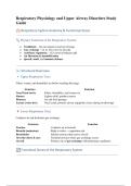
Respiratory Physiology and Upper Airway Disorders Study
Guide
🫁 Respiratory System Anatomy & Functional Zones
🔍 Primary Functions of the Respiratory System
● Ventilation – Air movement in and out of lungs
● Gas exchange – O₂ in, CO₂ out (via alveoli)
● Acid-base regulation – CO₂ removal balances pH
● Air filtration & humidification
● Speech, smell, and immune defense
🌬️ Structural Overview
🔹 Upper Respiratory Tract
Filters, warms, and humidifies air before reaching the lungs
Structure Function
Nose/Nasal cavity Filters, humidifies, and warms air
Sinuses Lighten skull, produce mucus
Pharynx Air and food passage
Larynx (voice box) Vocal cords, protects airway (epiglottis closes during swallowing)
🔻 Lower Respiratory Tract
Conducts air and facilitates gas exchange
Structure Function
Trachea Conducts air to bronchi
Bronchi (mainstem) Right is wider → aspiration risk
Bronchioles Smaller airways that lead to alveoli
Alveolar ducts & sacs Terminal airways where gas exchange occurs
Alveoli Primary site of gas exchange with pulmonary capillaries
🔄 Functional Zones of the Respiratory System
,🔹 1. Conducting Zone
Function: Move air in/out — no gas exchange
Nose → Nasal cavity → Pharynx → Larynx → Trachea → Bronchi → Bronchioles →
Includes
Terminal bronchioles
Lined with Ciliated epithelium + goblet cells → mucociliary escalator
Purpose Clean, humidify, and warm incoming air
🔻 2. Respiratory Zone
Function: Site of gas exchange
Includes Respiratory bronchioles → Alveolar ducts → Alveolar sacs → Alveoli
Walls Extremely thin; surrounded by pulmonary capillaries
Key cells Type I = gas exchange, Type II = surfactant production
Gas movement O₂ diffuses into blood, CO₂ diffuses out to alveoli
📊 Comparison: Conducting vs Respiratory Zones
Feature Conducting Zone Respiratory Zone
❌ ✅
Function Air transport Gas exchange
Gas exchange? No Yes
Epithelium Ciliated columnar + mucus Simple squamous
Ends at Terminal bronchioles Alveoli
🫁 Lungs & Supporting Structures
Structure Description
Right lung 3 lobes: upper, middle, lower
Left lung 2 lobes: upper, lower (cardiac notch)
Pleura Double membrane (visceral & parietal) with lubricating pleural fluid
Diaphragm Main muscle of inspiration, innervated by phrenic nerve (C3–C5)
🧪 Clinical Relevance
● Aspiration → more likely in right main bronchus
● Atelectasis → collapse of alveoli (↓ surfactant or poor inflation)
● Pneumonia → affects respiratory zone, impairs gas exchange
● Asthma, Bronchitis → affect conducting zone, cause airflow obstruction
● Emphysema → damages alveoli, ↓ surface area for diffusion
,🫀 Pulmonary Circulation & Gas Exchange
🔄 Overview
Pulmonary circulation is the low-pressure vascular system responsible for carrying
deoxygenated blood from the right heart to the lungs and returning oxygenated blood to the
left heart.
🔁 Pathway of Pulmonary Circulation
1. Right ventricle
2. → Pulmonary artery (only artery carrying deoxygenated blood)
3. → Pulmonary arterioles
4. → Pulmonary capillaries (surround alveoli — site of gas exchange)
5. → Pulmonary venules and veins
6. → Left atrium (now oxygen-rich)
🧬 Gas Exchange at the Alveolar-Capillary Membrane
✅ Occurs via diffusion
● O₂ moves from alveoli → pulmonary capillaries
● CO₂ moves from capillaries → alveoli to be exhaled
✅ Requires:
● Ventilation: Airflow into alveoli
● Perfusion: Blood flow through pulmonary capillaries
● Intact alveolar-capillary membrane
🧠 Efficiency of gas exchange depends on:
● Surface area of alveoli (↓ in emphysema)
● Thickness of membrane (↑ in fibrosis, pulmonary edema)
● Matching of ventilation and perfusion (V/Q ratio)
, 📊 Ventilation-Perfusion (V/Q) Ratio
● Normal V/Q ratio = ~0.8
o Ventilation (V) ≈ 4 L/min
o Perfusion (Q) ≈ 5 L/min
Abnormality Description Examples
Low V/Q (shunt) Perfusion > ventilation Atelectasis, pneumonia, mucus plug
High V/Q (dead space) Ventilation > perfusion Pulmonary embolism
True Shunt No ventilation at all ARDS, alveolar collapse
🧪 Clinical Relevance
● Hypoxemia can result from:
o V/Q mismatch
o Shunting
o Diffusion defects (fibrosis, edema)
● Pulmonary embolism = perfusion problem → high V/Q
● Pneumonia/atelectasis = ventilation problem → low V/Q
● ARDS = shunt physiology → no gas exchange despite oxygen therapy
👩⚕️ Nursing Implications
● Position patient to optimize V/Q (good lung down)
● Administer O₂ to improve PaO₂
● Encourage coughing, deep breathing, incentive spirometry
● Recognize signs of poor oxygenation: restlessness, tachypnea, ↓ SpO₂
🫁 Causes of Hypoxemia
(Low oxygen in arterial blood — PaO₂ < 80 mmHg)
📊 5 Major Mechanisms of Hypoxemia
Mechanism Definition Examples Key ABG Finding
CNS depression (opioids, head
↓ Air movement → ↓ O₂ in ↑ PaCO₂, ↓ PaO₂
1. Hypoventilation injury), neuromuscular disease,
alveoli (respiratory acidosis)
obesity
Guide
🫁 Respiratory System Anatomy & Functional Zones
🔍 Primary Functions of the Respiratory System
● Ventilation – Air movement in and out of lungs
● Gas exchange – O₂ in, CO₂ out (via alveoli)
● Acid-base regulation – CO₂ removal balances pH
● Air filtration & humidification
● Speech, smell, and immune defense
🌬️ Structural Overview
🔹 Upper Respiratory Tract
Filters, warms, and humidifies air before reaching the lungs
Structure Function
Nose/Nasal cavity Filters, humidifies, and warms air
Sinuses Lighten skull, produce mucus
Pharynx Air and food passage
Larynx (voice box) Vocal cords, protects airway (epiglottis closes during swallowing)
🔻 Lower Respiratory Tract
Conducts air and facilitates gas exchange
Structure Function
Trachea Conducts air to bronchi
Bronchi (mainstem) Right is wider → aspiration risk
Bronchioles Smaller airways that lead to alveoli
Alveolar ducts & sacs Terminal airways where gas exchange occurs
Alveoli Primary site of gas exchange with pulmonary capillaries
🔄 Functional Zones of the Respiratory System
,🔹 1. Conducting Zone
Function: Move air in/out — no gas exchange
Nose → Nasal cavity → Pharynx → Larynx → Trachea → Bronchi → Bronchioles →
Includes
Terminal bronchioles
Lined with Ciliated epithelium + goblet cells → mucociliary escalator
Purpose Clean, humidify, and warm incoming air
🔻 2. Respiratory Zone
Function: Site of gas exchange
Includes Respiratory bronchioles → Alveolar ducts → Alveolar sacs → Alveoli
Walls Extremely thin; surrounded by pulmonary capillaries
Key cells Type I = gas exchange, Type II = surfactant production
Gas movement O₂ diffuses into blood, CO₂ diffuses out to alveoli
📊 Comparison: Conducting vs Respiratory Zones
Feature Conducting Zone Respiratory Zone
❌ ✅
Function Air transport Gas exchange
Gas exchange? No Yes
Epithelium Ciliated columnar + mucus Simple squamous
Ends at Terminal bronchioles Alveoli
🫁 Lungs & Supporting Structures
Structure Description
Right lung 3 lobes: upper, middle, lower
Left lung 2 lobes: upper, lower (cardiac notch)
Pleura Double membrane (visceral & parietal) with lubricating pleural fluid
Diaphragm Main muscle of inspiration, innervated by phrenic nerve (C3–C5)
🧪 Clinical Relevance
● Aspiration → more likely in right main bronchus
● Atelectasis → collapse of alveoli (↓ surfactant or poor inflation)
● Pneumonia → affects respiratory zone, impairs gas exchange
● Asthma, Bronchitis → affect conducting zone, cause airflow obstruction
● Emphysema → damages alveoli, ↓ surface area for diffusion
,🫀 Pulmonary Circulation & Gas Exchange
🔄 Overview
Pulmonary circulation is the low-pressure vascular system responsible for carrying
deoxygenated blood from the right heart to the lungs and returning oxygenated blood to the
left heart.
🔁 Pathway of Pulmonary Circulation
1. Right ventricle
2. → Pulmonary artery (only artery carrying deoxygenated blood)
3. → Pulmonary arterioles
4. → Pulmonary capillaries (surround alveoli — site of gas exchange)
5. → Pulmonary venules and veins
6. → Left atrium (now oxygen-rich)
🧬 Gas Exchange at the Alveolar-Capillary Membrane
✅ Occurs via diffusion
● O₂ moves from alveoli → pulmonary capillaries
● CO₂ moves from capillaries → alveoli to be exhaled
✅ Requires:
● Ventilation: Airflow into alveoli
● Perfusion: Blood flow through pulmonary capillaries
● Intact alveolar-capillary membrane
🧠 Efficiency of gas exchange depends on:
● Surface area of alveoli (↓ in emphysema)
● Thickness of membrane (↑ in fibrosis, pulmonary edema)
● Matching of ventilation and perfusion (V/Q ratio)
, 📊 Ventilation-Perfusion (V/Q) Ratio
● Normal V/Q ratio = ~0.8
o Ventilation (V) ≈ 4 L/min
o Perfusion (Q) ≈ 5 L/min
Abnormality Description Examples
Low V/Q (shunt) Perfusion > ventilation Atelectasis, pneumonia, mucus plug
High V/Q (dead space) Ventilation > perfusion Pulmonary embolism
True Shunt No ventilation at all ARDS, alveolar collapse
🧪 Clinical Relevance
● Hypoxemia can result from:
o V/Q mismatch
o Shunting
o Diffusion defects (fibrosis, edema)
● Pulmonary embolism = perfusion problem → high V/Q
● Pneumonia/atelectasis = ventilation problem → low V/Q
● ARDS = shunt physiology → no gas exchange despite oxygen therapy
👩⚕️ Nursing Implications
● Position patient to optimize V/Q (good lung down)
● Administer O₂ to improve PaO₂
● Encourage coughing, deep breathing, incentive spirometry
● Recognize signs of poor oxygenation: restlessness, tachypnea, ↓ SpO₂
🫁 Causes of Hypoxemia
(Low oxygen in arterial blood — PaO₂ < 80 mmHg)
📊 5 Major Mechanisms of Hypoxemia
Mechanism Definition Examples Key ABG Finding
CNS depression (opioids, head
↓ Air movement → ↓ O₂ in ↑ PaCO₂, ↓ PaO₂
1. Hypoventilation injury), neuromuscular disease,
alveoli (respiratory acidosis)
obesity


