Radiology
Introduction(+everything you need to know about breast imaging):
X-Rays was discovered by Wilhelm Conrad Röntgen on 8 November 1895.
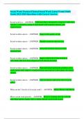
Electromagnetic spectrum:
Photons that propagate in vacuum in straight lines at the speed of light;
(Gamma ray → X-ray → Ultraviolet → Visible light → Infrared → Microwaves → Radio-waves)
Wave length
Photon energy (Kev) & Frequency
X-rays:
Uses:
1. X-ray crystallography. 3. CT scan.
2. Mammography. 4. Airport security.
Produced by energy conversion in the X-ray tube.
A diagnostic image is composed of differences in contrast between tissues which result from
differences in radiation interaction in the tissues; Absorption1, Transmission2, Scattering3.
Tissue appearances on x-ray:
1. Air → Black.
2. Fat → Dark grey.
3. Soft tissues → Grey
4. Bone, calcium → White.
5. Metal → Very white.
Mammography:
Mammography is a special type of X-ray imaging used to create detailed imaging of the breast.
Low-Kev X-ray.
High contrast.
High-resolution film.
X-ray system designed specifically for imaging the breasts.
Mortality risk from mammography induced radiation is 5 deaths/ million pts, using screen
film mammography. Sensitivity in women > 50;
1 2
Types: screening mammogram , diagnostic mammogram . 98% fatty breast.
Goals: 84% dense breasts.
1. High sensitivity for disease & Low false negative rate.
2. Lower specificity acceptable; 82-98%.
, Breast Cancer, why to Screen?
1. High prevalence
2. Improved outcome by treatment during the asymptomatic period.
3. Significant impact on public health.
4. Mortality reduction; Due to detection of cancers at smaller size/earlier stage;
Mammographically the lump could be visible 3-5 years before being palpable.
5. Increased detection of DCIS.
6. Early stage disease is curable.
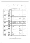
Screening Mammography Basically obtained views are:
1. Mediolateral-oblique (MLO).
Pectoralis major muscle is seen.
Upper portion / lower portion are used to describe lesion location.
2. Cranial-Caudal (CC).
Lateral (outer) portion & medial (inner) portion.
Additional views maybe obtained for further clarification; compression1, magnification2;
examples: Summation of fibro-glandular tissue1, Hidden masses2, Calcifications3.
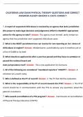
While screening we search for:
1. Masses:
Benign masses:
i. Tend to be spherical with smooth borders.
ii. If they contain calcification, it is macro, punctuate & Similar density than
that seen in carcinoma.
Malignant lesions:
i. Tend to be of variable shape, irregular outline.
ii. Calcification; different particle shape & density & the cluster shape is
irregular or triangular pointing toward the nipple).
iii. Speculated Dense mass.
iv. Architectural distortion.
v. Asymmetry of breast tissue.
vi. Skin thickening.
vii. Pathological lymph nodes; benign are rounded, well defined with fatty centre
(lucent center) while pathological lymph node are dense & opaque (no fatty
center).
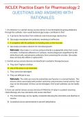
, 2. Calcifications:
Most are benign and can be dismissed.
The goal is to identify new or increasing calcifications or those with suspicious
morphology (Cluster of linear & branching micro-calcifications).
Benign calcifications:
1. Skin or dermal calcifications.
2. Vascular calcifications.
3. Lucent-centered calcifications (Fat necrosis).
4. Egg-shell or rim calcifications (Fat necrosis or calcification in cyst wall).
5. Coarse or popcorn calcification (Fibro adenoma).
6. Secretory calcifications, large rod like calcifications.
7. Punctuate calcification (less than 0.5mm).
8. Milk of calcium (in dependant part of the cyst).
9. Suture calcification.
10. Dystrophic calcifications (Trauma, surgery and irradiation).
ACS Screening Guidelines (Average Risk):
1. Annual mammography age 40 and older.
In Jordan every 2 years for asymptomatic patient and her age is more than 40.
Reduction in mortality by 30-50%
2. Annual clinical breast exam age 40 and older (Q 3 years age 20-40).
3. Self breast exam optional.
4. Annual mammography earlier if mother or sister diagnosed with breast cancer.
5. MRI if at high risk for breast cancer.
5 – 15% of breast cancers are not detected Mammographically;
1. Occult on mammogram (lobular CA).
2. Finding obscured by dense tissue.
3. Technical Error of interpretation.
On the mammogram report:
BI-RADS CODING
CATEGORY 0 Incomplete; needs further evaluation.
CATEGORY 1 Negative mammogram.
Introduction(+everything you need to know about breast imaging):
X-Rays was discovered by Wilhelm Conrad Röntgen on 8 November 1895.
Electromagnetic spectrum:
Photons that propagate in vacuum in straight lines at the speed of light;
(Gamma ray → X-ray → Ultraviolet → Visible light → Infrared → Microwaves → Radio-waves)
Wave length
Photon energy (Kev) & Frequency
X-rays:
Uses:
1. X-ray crystallography. 3. CT scan.
2. Mammography. 4. Airport security.
Produced by energy conversion in the X-ray tube.
A diagnostic image is composed of differences in contrast between tissues which result from
differences in radiation interaction in the tissues; Absorption1, Transmission2, Scattering3.
Tissue appearances on x-ray:
1. Air → Black.
2. Fat → Dark grey.
3. Soft tissues → Grey
4. Bone, calcium → White.
5. Metal → Very white.
Mammography:
Mammography is a special type of X-ray imaging used to create detailed imaging of the breast.
Low-Kev X-ray.
High contrast.
High-resolution film.
X-ray system designed specifically for imaging the breasts.
Mortality risk from mammography induced radiation is 5 deaths/ million pts, using screen
film mammography. Sensitivity in women > 50;
1 2
Types: screening mammogram , diagnostic mammogram . 98% fatty breast.
Goals: 84% dense breasts.
1. High sensitivity for disease & Low false negative rate.
2. Lower specificity acceptable; 82-98%.
, Breast Cancer, why to Screen?
1. High prevalence
2. Improved outcome by treatment during the asymptomatic period.
3. Significant impact on public health.
4. Mortality reduction; Due to detection of cancers at smaller size/earlier stage;
Mammographically the lump could be visible 3-5 years before being palpable.
5. Increased detection of DCIS.
6. Early stage disease is curable.
Screening Mammography Basically obtained views are:
1. Mediolateral-oblique (MLO).
Pectoralis major muscle is seen.
Upper portion / lower portion are used to describe lesion location.
2. Cranial-Caudal (CC).
Lateral (outer) portion & medial (inner) portion.
Additional views maybe obtained for further clarification; compression1, magnification2;
examples: Summation of fibro-glandular tissue1, Hidden masses2, Calcifications3.
While screening we search for:
1. Masses:
Benign masses:
i. Tend to be spherical with smooth borders.
ii. If they contain calcification, it is macro, punctuate & Similar density than
that seen in carcinoma.
Malignant lesions:
i. Tend to be of variable shape, irregular outline.
ii. Calcification; different particle shape & density & the cluster shape is
irregular or triangular pointing toward the nipple).
iii. Speculated Dense mass.
iv. Architectural distortion.
v. Asymmetry of breast tissue.
vi. Skin thickening.
vii. Pathological lymph nodes; benign are rounded, well defined with fatty centre
(lucent center) while pathological lymph node are dense & opaque (no fatty
center).
, 2. Calcifications:
Most are benign and can be dismissed.
The goal is to identify new or increasing calcifications or those with suspicious
morphology (Cluster of linear & branching micro-calcifications).
Benign calcifications:
1. Skin or dermal calcifications.
2. Vascular calcifications.
3. Lucent-centered calcifications (Fat necrosis).
4. Egg-shell or rim calcifications (Fat necrosis or calcification in cyst wall).
5. Coarse or popcorn calcification (Fibro adenoma).
6. Secretory calcifications, large rod like calcifications.
7. Punctuate calcification (less than 0.5mm).
8. Milk of calcium (in dependant part of the cyst).
9. Suture calcification.
10. Dystrophic calcifications (Trauma, surgery and irradiation).
ACS Screening Guidelines (Average Risk):
1. Annual mammography age 40 and older.
In Jordan every 2 years for asymptomatic patient and her age is more than 40.
Reduction in mortality by 30-50%
2. Annual clinical breast exam age 40 and older (Q 3 years age 20-40).
3. Self breast exam optional.
4. Annual mammography earlier if mother or sister diagnosed with breast cancer.
5. MRI if at high risk for breast cancer.
5 – 15% of breast cancers are not detected Mammographically;
1. Occult on mammogram (lobular CA).
2. Finding obscured by dense tissue.
3. Technical Error of interpretation.
On the mammogram report:
BI-RADS CODING
CATEGORY 0 Incomplete; needs further evaluation.
CATEGORY 1 Negative mammogram.