PAS 407 Nerves and Neuropathy UPDATED ACTUAL Questions and CORRECT
Answers
partial/complete damage to peripheral nerves
peripheral neuropathies
- regional losses of sensory and motor function
- physical trauma (laceration, mechanical compres-
sion/crush, traction)
- chemical (alcohol, medication)
what are causes of peripheral neuropathies?
- metabolic (diabetes mellitus, hypothyroidism)
- genetic/other diseases (Charcot-Marie-Tooth diseases,
Guillain-Barre syndrome)
occurs days to weeks
period in the weeks following traumatic neural leison
acute musculoskeletal neuropathy
characterized by loss of sensation and motor function
- no functional deformity (normal muscle bulk, joint archi-
tecture)
occurs months to years (long term deinnervation)
can be
chronic musculoskeletal neuropathy
- progressive degeneration over weeks/months (e.g. dia-
betic neuropathy, compartment syndrome)
- unresolved acute neural lesion
- muscle wasting (loss in muscle bulk)
- joint contracture (unopposed antagonistic muscle pull
what physical deformities are accompanied to weakness
alters joint architecture)
in chronic neuropathy?
note: contracture keeps joints from moving freely
- cutaneous changes (shiny, brittle skin, loss of hair)
, derived from lateral cord of brachial plexus (C5-C7)
and innervates to coracobrachialis, biceps brachii, and
musculocutaneous nerve
brachialis muscles, terminating as the lateral cutaneous
nerve of forearm
weakness of arm flexion and sensory loss along the lateral
forearm
injuries to musculocutaneous nerve
note: musculocutaneous innervates to anterior compart-
ment muscles, therefore complete arm flexion
traction injury to upper brachial nerve
results from combination of excessive contralateral neck
brachial plexus nerve leisons (Erb-Duchenne nerve palsy)
flexion with shoulder depression
- fall on shoulder
- diflcult labor
results in "waiter's tip" position
- internal shoulder rotation
- elbow extension
Erb-Duchenne Palsy - forearm pronated
- wrist flexed
C5-T1
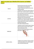
enters forearm by passing heads of pronator teres
median nerve passes within neurovascular plane between flexor digito-
rum superficialis and profundus
continues distally lateral to flexor digitorum superficialis
Answers
partial/complete damage to peripheral nerves
peripheral neuropathies
- regional losses of sensory and motor function
- physical trauma (laceration, mechanical compres-
sion/crush, traction)
- chemical (alcohol, medication)
what are causes of peripheral neuropathies?
- metabolic (diabetes mellitus, hypothyroidism)
- genetic/other diseases (Charcot-Marie-Tooth diseases,
Guillain-Barre syndrome)
occurs days to weeks
period in the weeks following traumatic neural leison
acute musculoskeletal neuropathy
characterized by loss of sensation and motor function
- no functional deformity (normal muscle bulk, joint archi-
tecture)
occurs months to years (long term deinnervation)
can be
chronic musculoskeletal neuropathy
- progressive degeneration over weeks/months (e.g. dia-
betic neuropathy, compartment syndrome)
- unresolved acute neural lesion
- muscle wasting (loss in muscle bulk)
- joint contracture (unopposed antagonistic muscle pull
what physical deformities are accompanied to weakness
alters joint architecture)
in chronic neuropathy?
note: contracture keeps joints from moving freely
- cutaneous changes (shiny, brittle skin, loss of hair)
, derived from lateral cord of brachial plexus (C5-C7)
and innervates to coracobrachialis, biceps brachii, and
musculocutaneous nerve
brachialis muscles, terminating as the lateral cutaneous
nerve of forearm
weakness of arm flexion and sensory loss along the lateral
forearm
injuries to musculocutaneous nerve
note: musculocutaneous innervates to anterior compart-
ment muscles, therefore complete arm flexion
traction injury to upper brachial nerve
results from combination of excessive contralateral neck
brachial plexus nerve leisons (Erb-Duchenne nerve palsy)
flexion with shoulder depression
- fall on shoulder
- diflcult labor
results in "waiter's tip" position
- internal shoulder rotation
- elbow extension
Erb-Duchenne Palsy - forearm pronated
- wrist flexed
C5-T1
enters forearm by passing heads of pronator teres
median nerve passes within neurovascular plane between flexor digito-
rum superficialis and profundus
continues distally lateral to flexor digitorum superficialis