Exam 1 Study Guide
Week 1 Lab: Outcome Measures -
When to perform outcome measures
- When the Pt first comes in (evaluation), re-eval, and last day of therapy
Why we use outcome measures in outpatient therapy
- Establishes baseline performance and tracks progress
- Supports reimbursement and need for skilled therapy
- Often mandated by insurance
- They are standardized assessments
DASH score: is higher or lower score better (which score means LESS function)?
- Most common outcome measure used in outpatient OT
- lower score is better = more function
- higher score = less function
How to administer the quick DASH
- Pt must answer every question
- Questions are based off Pt’s condition in the last week
- Pt gives best estimate if they have not performed the activity
- Questions are answered based on Pt’s ability to use both hands, not just the injured side
Week 2: Wound Healing/Scar Management -
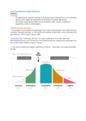
Phases of wound healing: purpose of each phase and how long each phase lasts
- Hemostasis: blood vessels open to bring oxygen and nutrients to the wound
- Inflammatory phase (week 1): WBCs and macrophages migrate into the wound to clean up.
Clotting occurs
o Stitches in
- Proliferative/fibroblastic phase (week 2-3): wound closes and scar synthesis begins. Collagen
laid down
o Things are healing
o Therapy typically starts here
- Maturation/remodeling phase (week 3 – 1 year): collagen matrix is remodeling and the tissue
changes over time
Types of wound closure and pro’s/con’s of each
- Primary: MOST COMMON
o Typically post-surgery sutures
o Closure needs to be don’t quickly (4-6 hours after injury)
o Edges of wound are approximated and hed together with sutures, steri-strips, or
surgical adhesive
o Wound bed is closed and covered with skin
, o PRO: reduces risk of infection and scarring is limited
- Secondary:
o Very slow to heal
o Wound edges cannot be approximated, wound is left open to fill in from the bottom up
and sides inward
o Could occur if wound “springs open” after primary intention
o CON: larger scar and higher risk of infection
- Tertiary:
o Combination of primary and secondary intention
o Wound is initially left open to drain or be irrigated to prevent infection then wound is
closed via primary closure
o Examples: dog or animal bites, traumatic injuries (MVS, firework explosion) or injury in
dirty environment where debris might be present
Tensile strength timeline
- Tensile strength: the maximum amount of force applied to a soft tissue structure before it will
rupture
- Tensile strength increases at the collagen fibers → collagen gives skin strength
- Tensile strength is directly related to time in a wound
o Very slow for the first two – three weeks after injury
o Reaches a peak around 60 days (8 weeks) after injury but for some, can take several
months to reach maximum of 80% OF NORMAL TISSUE STRENGTH
- Tensile strength timeline:
o 4 weeks = 40-50%
o 6 weeks = 60%
o 8 weeks = 70-80% (WOUND STRONG ENOUGH TO BE AGGRESSIVE WITH THERAPY)
o Will NEVER be 100%
Wound classification terms
• Partial vs full thickness
o Partial: tissue injury the extends through the epidermis and partial dermis
▪ Heals by scabbing (epithelization) -→ new cells migrate across a wound for closure
▪ Examples: skin tears, abrasions, tape burns, blisters
o Full: tissue destruction extending through the epidermis and dermis into subcutaneous
tissue (fat, tendon, muscle, even bone)
▪ Healing process: granulation, contraction, and epithelization
▪ Clinical examples: surgical incisions, donor sites for grafting, third and fourth
degree burns, stage 3 and 4 ulcers
• Color
o Red: Healthy granulating tissue is dark pink or red
▪ TX: Focus on protection and wound closure
▪ Red around the wound or red streaking indicates possible infection!
o Yellow: Drainage and slough: yellow pus and dead debris → macrophages are responding
to inflammation
Week 1 Lab: Outcome Measures -
When to perform outcome measures
- When the Pt first comes in (evaluation), re-eval, and last day of therapy
Why we use outcome measures in outpatient therapy
- Establishes baseline performance and tracks progress
- Supports reimbursement and need for skilled therapy
- Often mandated by insurance
- They are standardized assessments
DASH score: is higher or lower score better (which score means LESS function)?
- Most common outcome measure used in outpatient OT
- lower score is better = more function
- higher score = less function
How to administer the quick DASH
- Pt must answer every question
- Questions are based off Pt’s condition in the last week
- Pt gives best estimate if they have not performed the activity
- Questions are answered based on Pt’s ability to use both hands, not just the injured side
Week 2: Wound Healing/Scar Management -
Phases of wound healing: purpose of each phase and how long each phase lasts
- Hemostasis: blood vessels open to bring oxygen and nutrients to the wound
- Inflammatory phase (week 1): WBCs and macrophages migrate into the wound to clean up.
Clotting occurs
o Stitches in
- Proliferative/fibroblastic phase (week 2-3): wound closes and scar synthesis begins. Collagen
laid down
o Things are healing
o Therapy typically starts here
- Maturation/remodeling phase (week 3 – 1 year): collagen matrix is remodeling and the tissue
changes over time
Types of wound closure and pro’s/con’s of each
- Primary: MOST COMMON
o Typically post-surgery sutures
o Closure needs to be don’t quickly (4-6 hours after injury)
o Edges of wound are approximated and hed together with sutures, steri-strips, or
surgical adhesive
o Wound bed is closed and covered with skin
, o PRO: reduces risk of infection and scarring is limited
- Secondary:
o Very slow to heal
o Wound edges cannot be approximated, wound is left open to fill in from the bottom up
and sides inward
o Could occur if wound “springs open” after primary intention
o CON: larger scar and higher risk of infection
- Tertiary:
o Combination of primary and secondary intention
o Wound is initially left open to drain or be irrigated to prevent infection then wound is
closed via primary closure
o Examples: dog or animal bites, traumatic injuries (MVS, firework explosion) or injury in
dirty environment where debris might be present
Tensile strength timeline
- Tensile strength: the maximum amount of force applied to a soft tissue structure before it will
rupture
- Tensile strength increases at the collagen fibers → collagen gives skin strength
- Tensile strength is directly related to time in a wound
o Very slow for the first two – three weeks after injury
o Reaches a peak around 60 days (8 weeks) after injury but for some, can take several
months to reach maximum of 80% OF NORMAL TISSUE STRENGTH
- Tensile strength timeline:
o 4 weeks = 40-50%
o 6 weeks = 60%
o 8 weeks = 70-80% (WOUND STRONG ENOUGH TO BE AGGRESSIVE WITH THERAPY)
o Will NEVER be 100%
Wound classification terms
• Partial vs full thickness
o Partial: tissue injury the extends through the epidermis and partial dermis
▪ Heals by scabbing (epithelization) -→ new cells migrate across a wound for closure
▪ Examples: skin tears, abrasions, tape burns, blisters
o Full: tissue destruction extending through the epidermis and dermis into subcutaneous
tissue (fat, tendon, muscle, even bone)
▪ Healing process: granulation, contraction, and epithelization
▪ Clinical examples: surgical incisions, donor sites for grafting, third and fourth
degree burns, stage 3 and 4 ulcers
• Color
o Red: Healthy granulating tissue is dark pink or red
▪ TX: Focus on protection and wound closure
▪ Red around the wound or red streaking indicates possible infection!
o Yellow: Drainage and slough: yellow pus and dead debris → macrophages are responding
to inflammation