know it !
· me ⑨
⑤ on there !!
· SH review
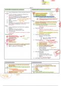
UNIT 3
ACUTE BRO ↑ compliance & ↓ resistance
● Acute inflammation of the trachea & bronchi
CHRONIC BRO ↑compliance ↓ resistance
Causes: "repeated
*
Causes:
● Viral: Flu viruses, coronavirus, rhinovirus, ⑳● Cigarette smoking (90%)**
● Genetic predisposition
airway
Functions
adenovirus, coxsackievirus D
● Inhalation of irritants
● Nonviral: Streptococcus PNA, haemophilus ● Referred “Type B” COPD *BLUE BLOATER
influenzae, mycoplasma, moraxella, & ○ Hypersecretion of bronchial mucus
chlamydia PNA
● Heat #recent onset of cough
temp
-○ Chronic or recurrent prod cough >3m &
occurring >2+ yrs
● Allergic reactions - low grade Patho:
● Smokers + sore throat ● Chronic inflammation & swelling of bronchial 1
● Swelling of bronchial mucosa in kids ass w/
resp distress, wheezing is “asthmatic D
mucosa → to scarring loss of clary function
>
-
● ↑ mucus prod w/ formation of mucus plugs reque
Any
bronchitis” ● ↑ bronchial wall thickness
Patho: ○ Resistance ↑ work of breathing & O2 =
● Airway inflamed & narrowed from capillary demands
dilation ● Pulmonary HTN**
● Swelling from fluid exudation ○ Inflamm in bronchial walls w/
● Infiltration w/ inflammatory cells vasoconstriction of pulmonary vessels &
● ↑ mucus prod arteries
● Loss of ciliary function & portions of ciliated
epithelium
O ○ R side HF may occur r/t high pulmonary
resistance** "Cor pulmonace"
Manifestations: ● Destruction of bronchial walls
● Mild & self limiting onset ○ Dilation of airway space
* ● Cough (prod or non) cough ○ Causes:
-
● Low grade fever trachea + bronchi ■ Infec from STREP, STAPH PNA
● Chest discomfort inflamm ■ Infection w/ mold
o ● Sore throat ● Manifestations: *
44y0 overweight
& ● Fatigue ○ >30 to 40 yrs -
DX:
● Postnasal drip -
○ 1:2 male to female ratio *overweight*
-
⑨○ SOB on exertion
① ● Hallmark onset distinct cough (spasmodic)** ○ Lots of sputum** "purulent"
● Chest x-ray to distinguish from PNA D○ Chronic cough (severe in AM)**
● WBC or sputum is not reliable ○ Excess body fluids (edema,
hypervolemia)** + polycythemia
○ Hx of smoking
○ Chills, malaise, muscle aches, fatigue, loss
·
of libido & insomnia
● END STAGE signs:
○ R side HF
○ Distended neck veins
○ R ventricular heave & gallo
○ Edema
○ HTN, Cyanosis = late sign
● DX: Chest x-ray, ECG, PFT, ABGs
HYPOVENTILATION HYPERVENTILATION
● Air to alveoli is insufficient to provide O2 & ● ↑ in amount of air entering alveoli
remove CO2 ● Leads to hypocapnia (PACO2 < 35 mm Hg)
⑧● Results in ↑ PACO2 (>45mm Hg) & hypoxemia More common causes: ↳ Fr alkalos is
Causes: ● Pain depth
↑ RR and
● **Drugs: Morphine ● Fever
○ Barbiturates (depresses CRD) ● Anxiety
● Obesity (Pickwickian syndrome)
T
Less common causes:
● Myasthenia gravis + COPD ⑨● Obstructive & restrictive lung disease
rhyp me
⑫
*
, ● Obstructive sleep apnea ● Sepsis
● Chest wall damage ● Brainstem injury
● Paralysis of resp muscles ①
● High altitude**
response
● Pain r/t sx of thorax or abdomen
↳ normal comp
.
○ 2ndary to ↓ aspiration
HYPOXEMIA HYPOXIA
● Deficient levels of blood oxygen as measured
* circulatory hypoxia-cardiac
● ↓ in tissue oxygenation
arrest, cyanosissentation
by low arterial O2 concentration & low Types: & cardiac
↳
Hemoglobin saturation as measured by Hypoxic hypoxia output
arterial blood gases or pulse ox ● PaO2 is ↓ despite normal O2 capacity
*KEY POINTS PG 478* ○ Causes: high altitude, hypoventilation &
● O2 & CO2 diffuse quickly across alveolar airway obstruction
capillary membranes Anemic hypoxia
● O2 is carried in 2 forms: ● Low Hgb
○ Dissolved in solution Circulatory hypoxia
○ Bound to hgb ● Low cardiac output (O2 carrying capacity is
● More O2 is bound than dissolved normal but blood flow is reduced)
● Low hgb levels & sat affects the O2 content in ○ Shock, cardiac arrest, HF
blood Histotoxic hypoxia
● ↓ O2 carrying capacity from toxic substance
“Cyanide poisoning”
EMPHYSEMA Type A COPD “Pink Puffer”
Causes: def
alphatype
● Abnormal enlargement of the distal air sacs
D ● IRREVERSIBLE
● Smoking, Air pollution
● Certain occupations → welding, mining asbestos 1 .2 ,5 ,
4
● Smoking >70 packs/yr question
Patho: ⑭
⑧ ● Release of proteolytic enzymes from neutrophils & macrophages** to
Pa02
● Smoking causes alveolar damage in 2 ways: ↳ damage clubbing
a- alveoli
desitution
the up
○ 1. Leads to inflamm in lung tissue (parenchyma) porsed
wall
" -
○ 2. Inactivates a1-antitrypsin (protects the lung parenchyma)** underweight
⑨
-
● Loss of elastic tissue → ↓ in size of smaller bronchioles
● Loss of alveolar walls leads to reduc in pulmonary capillary bed
○ & leads to bullae (large, thin walled cysts in the lung)
Manifestations:
● Progressive exertional dyspnea
● Thin, hunched forward w/ SOB for 3-4 yrs
○ b/c work of ↑ resp effort & ↓ ability to consume calories
● ↑ in women who smoke
● Cough (minimal or absent)
● Prolonged expiration** "thin appearance"
See
Aris
~
● Digital clubbing** + PaO2 < 80
● Barrel chest**
● ↓ breath sounds, lack of crackles & rhonchi
● Wheezing
● Hyperresonance I
● Pursed lip breathing**
● LATE = dyspnea on exertion
SARCOIDOSIS *autoimmune* Common in women 30s-40s
Causes:
* rib cage will stiffen
WVONG "My
:
, abnormation
s
-N
①
● Acute or chronic systemic disease *unknown*
● 1st degree relative ↑ risk disease 5 fold**
Patho:
● Dev. of multiple uniform, noncaseating epithelioid granulomas**
● Affects lymphs nodes & lung tissue
⑨ ● Abnormal T-cells
granulomas
Manifestations:
S
-
● Mailasie, Fatigue, Fever
● Weight loss *choosing b/t eating & breathing* t
mentation
hyperpig
● Chest discomfort
● Dyspnea of insidious onset**
⑧ ● Dry, non prod cough**
⑨ ● Erythema nodosum (painful nodes)**
● Macles, papules, Hyperpigmentation**, subQ nodules
● Hepatosplenomegaly, lymphadenopathy
DX:
● Elevated liver enzymes (AST, ALT), Hypercalemia
● Chest x-ray, PFT
KYPHOSCOLIOSIS **A LOT of ON EXAM FROM THIS** ANKYLOSING SPONDYLITIS LOT of ON EXAM FROM
unknown
Causes: -> Causes:
-
·
● Idiopathic (80-90% cases) or congenital or ● More in males (3:1) ages 15-35
neuromuscular disease >
-muscular dystrophy ● Idiopathic
Patho: ● Chronic inflamm at site of ligamentous
● Bone deformity of chest wall results in insertion into spine
hunchback & scoliosis
● Higher deformity in vertebral column
● 90% have (+) HLA-B27 antigen
Patho:
limitedapansion
○ greater compromise of resp status** ● Immobility of the vertebral joints & fixation of
⑨● Lung volume compressed, leads to ribs
omprominen
atelectasis, mismatch, hypoxemia** ● Inflammatory affects the articular process,
Manifestations: costovertebral, sacroiliac joints
↳
·● Dyspnea on exertion ● Fibrotic response leads to joint calcification,
-my
● Rapid, shallow breathing ligament ossification & skeletal immobility** d
&
● Chest wall Manifestations:
&● Ribs protruding backward ● Low/middle back pain
○ flaring on convex side, crowded on ○ Stiffness w/ prolonged rest
concave side** ● w/ exercise = pain decrease
● Hypoxemia, CO2 retention = LATE ● Ribcage mvt reduced → restrictive lung
dysfunction**
● Chest wall muscular atrophy**
● Breathing prob d/t rib cage immobilization
deestos
HYPERSENSITIVITY PNEUMONITIS
Causes: >
-
● Extrinsic allergic alveolitis
⑧● Restrictive & occupational disease**
⑤● *PREDOMINANCE IN NONSMOKERS (80-95%)*
-
Patho:
● Antigen combines w/ serum antibody in alveolar walls
● Genetic predisposition**
● Leads to type 3 hypersensitivity reaction**
● Leads to diffuse pulmonary fibrosis in upper lobes**
· me ⑨
⑤ on there !!
· SH review
UNIT 3
ACUTE BRO ↑ compliance & ↓ resistance
● Acute inflammation of the trachea & bronchi
CHRONIC BRO ↑compliance ↓ resistance
Causes: "repeated
*
Causes:
● Viral: Flu viruses, coronavirus, rhinovirus, ⑳● Cigarette smoking (90%)**
● Genetic predisposition
airway
Functions
adenovirus, coxsackievirus D
● Inhalation of irritants
● Nonviral: Streptococcus PNA, haemophilus ● Referred “Type B” COPD *BLUE BLOATER
influenzae, mycoplasma, moraxella, & ○ Hypersecretion of bronchial mucus
chlamydia PNA
● Heat #recent onset of cough
temp
-○ Chronic or recurrent prod cough >3m &
occurring >2+ yrs
● Allergic reactions - low grade Patho:
● Smokers + sore throat ● Chronic inflammation & swelling of bronchial 1
● Swelling of bronchial mucosa in kids ass w/
resp distress, wheezing is “asthmatic D
mucosa → to scarring loss of clary function
>
-
● ↑ mucus prod w/ formation of mucus plugs reque
Any
bronchitis” ● ↑ bronchial wall thickness
Patho: ○ Resistance ↑ work of breathing & O2 =
● Airway inflamed & narrowed from capillary demands
dilation ● Pulmonary HTN**
● Swelling from fluid exudation ○ Inflamm in bronchial walls w/
● Infiltration w/ inflammatory cells vasoconstriction of pulmonary vessels &
● ↑ mucus prod arteries
● Loss of ciliary function & portions of ciliated
epithelium
O ○ R side HF may occur r/t high pulmonary
resistance** "Cor pulmonace"
Manifestations: ● Destruction of bronchial walls
● Mild & self limiting onset ○ Dilation of airway space
* ● Cough (prod or non) cough ○ Causes:
-
● Low grade fever trachea + bronchi ■ Infec from STREP, STAPH PNA
● Chest discomfort inflamm ■ Infection w/ mold
o ● Sore throat ● Manifestations: *
44y0 overweight
& ● Fatigue ○ >30 to 40 yrs -
DX:
● Postnasal drip -
○ 1:2 male to female ratio *overweight*
-
⑨○ SOB on exertion
① ● Hallmark onset distinct cough (spasmodic)** ○ Lots of sputum** "purulent"
● Chest x-ray to distinguish from PNA D○ Chronic cough (severe in AM)**
● WBC or sputum is not reliable ○ Excess body fluids (edema,
hypervolemia)** + polycythemia
○ Hx of smoking
○ Chills, malaise, muscle aches, fatigue, loss
·
of libido & insomnia
● END STAGE signs:
○ R side HF
○ Distended neck veins
○ R ventricular heave & gallo
○ Edema
○ HTN, Cyanosis = late sign
● DX: Chest x-ray, ECG, PFT, ABGs
HYPOVENTILATION HYPERVENTILATION
● Air to alveoli is insufficient to provide O2 & ● ↑ in amount of air entering alveoli
remove CO2 ● Leads to hypocapnia (PACO2 < 35 mm Hg)
⑧● Results in ↑ PACO2 (>45mm Hg) & hypoxemia More common causes: ↳ Fr alkalos is
Causes: ● Pain depth
↑ RR and
● **Drugs: Morphine ● Fever
○ Barbiturates (depresses CRD) ● Anxiety
● Obesity (Pickwickian syndrome)
T
Less common causes:
● Myasthenia gravis + COPD ⑨● Obstructive & restrictive lung disease
rhyp me
⑫
*
, ● Obstructive sleep apnea ● Sepsis
● Chest wall damage ● Brainstem injury
● Paralysis of resp muscles ①
● High altitude**
response
● Pain r/t sx of thorax or abdomen
↳ normal comp
.
○ 2ndary to ↓ aspiration
HYPOXEMIA HYPOXIA
● Deficient levels of blood oxygen as measured
* circulatory hypoxia-cardiac
● ↓ in tissue oxygenation
arrest, cyanosissentation
by low arterial O2 concentration & low Types: & cardiac
↳
Hemoglobin saturation as measured by Hypoxic hypoxia output
arterial blood gases or pulse ox ● PaO2 is ↓ despite normal O2 capacity
*KEY POINTS PG 478* ○ Causes: high altitude, hypoventilation &
● O2 & CO2 diffuse quickly across alveolar airway obstruction
capillary membranes Anemic hypoxia
● O2 is carried in 2 forms: ● Low Hgb
○ Dissolved in solution Circulatory hypoxia
○ Bound to hgb ● Low cardiac output (O2 carrying capacity is
● More O2 is bound than dissolved normal but blood flow is reduced)
● Low hgb levels & sat affects the O2 content in ○ Shock, cardiac arrest, HF
blood Histotoxic hypoxia
● ↓ O2 carrying capacity from toxic substance
“Cyanide poisoning”
EMPHYSEMA Type A COPD “Pink Puffer”
Causes: def
alphatype
● Abnormal enlargement of the distal air sacs
D ● IRREVERSIBLE
● Smoking, Air pollution
● Certain occupations → welding, mining asbestos 1 .2 ,5 ,
4
● Smoking >70 packs/yr question
Patho: ⑭
⑧ ● Release of proteolytic enzymes from neutrophils & macrophages** to
Pa02
● Smoking causes alveolar damage in 2 ways: ↳ damage clubbing
a- alveoli
desitution
the up
○ 1. Leads to inflamm in lung tissue (parenchyma) porsed
wall
" -
○ 2. Inactivates a1-antitrypsin (protects the lung parenchyma)** underweight
⑨
-
● Loss of elastic tissue → ↓ in size of smaller bronchioles
● Loss of alveolar walls leads to reduc in pulmonary capillary bed
○ & leads to bullae (large, thin walled cysts in the lung)
Manifestations:
● Progressive exertional dyspnea
● Thin, hunched forward w/ SOB for 3-4 yrs
○ b/c work of ↑ resp effort & ↓ ability to consume calories
● ↑ in women who smoke
● Cough (minimal or absent)
● Prolonged expiration** "thin appearance"
See
Aris
~
● Digital clubbing** + PaO2 < 80
● Barrel chest**
● ↓ breath sounds, lack of crackles & rhonchi
● Wheezing
● Hyperresonance I
● Pursed lip breathing**
● LATE = dyspnea on exertion
SARCOIDOSIS *autoimmune* Common in women 30s-40s
Causes:
* rib cage will stiffen
WVONG "My
:
, abnormation
s
-N
①
● Acute or chronic systemic disease *unknown*
● 1st degree relative ↑ risk disease 5 fold**
Patho:
● Dev. of multiple uniform, noncaseating epithelioid granulomas**
● Affects lymphs nodes & lung tissue
⑨ ● Abnormal T-cells
granulomas
Manifestations:
S
-
● Mailasie, Fatigue, Fever
● Weight loss *choosing b/t eating & breathing* t
mentation
hyperpig
● Chest discomfort
● Dyspnea of insidious onset**
⑧ ● Dry, non prod cough**
⑨ ● Erythema nodosum (painful nodes)**
● Macles, papules, Hyperpigmentation**, subQ nodules
● Hepatosplenomegaly, lymphadenopathy
DX:
● Elevated liver enzymes (AST, ALT), Hypercalemia
● Chest x-ray, PFT
KYPHOSCOLIOSIS **A LOT of ON EXAM FROM THIS** ANKYLOSING SPONDYLITIS LOT of ON EXAM FROM
unknown
Causes: -> Causes:
-
·
● Idiopathic (80-90% cases) or congenital or ● More in males (3:1) ages 15-35
neuromuscular disease >
-muscular dystrophy ● Idiopathic
Patho: ● Chronic inflamm at site of ligamentous
● Bone deformity of chest wall results in insertion into spine
hunchback & scoliosis
● Higher deformity in vertebral column
● 90% have (+) HLA-B27 antigen
Patho:
limitedapansion
○ greater compromise of resp status** ● Immobility of the vertebral joints & fixation of
⑨● Lung volume compressed, leads to ribs
omprominen
atelectasis, mismatch, hypoxemia** ● Inflammatory affects the articular process,
Manifestations: costovertebral, sacroiliac joints
↳
·● Dyspnea on exertion ● Fibrotic response leads to joint calcification,
-my
● Rapid, shallow breathing ligament ossification & skeletal immobility** d
&
● Chest wall Manifestations:
&● Ribs protruding backward ● Low/middle back pain
○ flaring on convex side, crowded on ○ Stiffness w/ prolonged rest
concave side** ● w/ exercise = pain decrease
● Hypoxemia, CO2 retention = LATE ● Ribcage mvt reduced → restrictive lung
dysfunction**
● Chest wall muscular atrophy**
● Breathing prob d/t rib cage immobilization
deestos
HYPERSENSITIVITY PNEUMONITIS
Causes: >
-
● Extrinsic allergic alveolitis
⑧● Restrictive & occupational disease**
⑤● *PREDOMINANCE IN NONSMOKERS (80-95%)*
-
Patho:
● Antigen combines w/ serum antibody in alveolar walls
● Genetic predisposition**
● Leads to type 3 hypersensitivity reaction**
● Leads to diffuse pulmonary fibrosis in upper lobes**