
Structure & Function of the Respiratory System
Sleep-related breathing disorders
Ventilation → movement of air in & out of the lungs (inspiration & expiration)
● With inspiration the diaphragm contracts
● With expiration diaphragm moves upward because it is relaxing
Perfusion → flow of blood through the lungs
Diffusion → transfer of gas between air-filled spaces of the lungs & the blood → crosses the alveolar/capillary
membrane. Affected by:
● Surface area available for diffusion → decreased in lung disease, lobectomy, pneumonectomy
● Thickness of alveolar capillary membrane → pneumonia, pulmonary edema
● Partial pressure of alveolar gasses → CO2 diffuses faster than O2
,Respiratory system structure and the conducting and respiratory airways
Lung structure
● Separated by the mediastinum & its contents
● Left lung = 2 lobes
● Right lung = 3 lobes
● Apex = upper part of the lung (lies against the top of the thoracic cavity)
● Base = lower part of the lung (lies against the diaphragm)
Respiratory system structure
● Nasopharynx, Oropharynx, and Larynx (connects the oropharynx to the trachea)
● The Trachea is supported by C-shaped rings of hyaline cartilage (divides into right and left main
bronchial at T5) → Has both vestibular folds & vocal folds that help produce speech & protect lungs
from substances other than air
● Carina (located between the main bronchi) has sensitive cells that cause violent coughing when
stimulated
● Tracheobronchial tree → trachea, bronchi and bronchioles
○ Trachea splits off into L & R bronchus → then to the bronchioles → then to the alveoli (where
gas exchange happens)
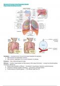
,Conducting airways → air moves as it passes between the atmosphere & the lungs
Respiratory airways → where gas exchange takes place
Bronchial and pulmonary circulations that supply the lungs
Pulmonary Circulation → comes from the pulmonary artery and provides for gas exchange. Oxygenated
blood leaves the right side of the heart and into the pulmonary artery. The pulmonary artery divides into left &
right. The pulmonary vein returns oxygenated blood to the left atrium
Bronchial Circulation → comes from the thoracic aorta and supplies the lungs with oxygen, distributes blood
to conducting airways, and warms & humidifies incoming air
● DOES NOT participate in gas exchange because it doesn’t pass through the alveolar capillaries (is
deoxygenated blood)
● Bronchial blood vessels form new blood vessels & can provide collateral circulation (example – if
someone has a pulmonary embolism and the pulmonary vessels in circulation are blocked…the
bronchial blood vessels develop a collateral circulation)
, Intrathoracic, intrapleural, and intra-alveolar pressures
Intrapulmonary (Alveolar) Pressure → pressure inside the airway & alveoli of the lungs
● During expiration pressure ↑ as the thoracic cavity volume decreases → when expiration stops the
pressure = atmospheric pressure
Intrapleural Pressure → pressure in the pleural cavity
● During inspiration, pressure drops causing ↓ intrathoracic airway pressure & airflow
Intrathoracic Pressure → pressure in the thoracic cavity = to the intrapleural pressure
● This is the pressure the heart, lungs, and great vessels are exposed to
● Ex → if someone is performing the valsalva maneuver unintentionally while pooping, intrathoracic
pressure increases; decreasing venous return to the heart
Lung compliance → the change in lung volume that can be achieved with a change in respiratory pressure
(takes more pressure to move air into noncompliant lungs than it does compliant lungs)
Airway resistance → volume of air that moves into & out of the air exchange portion of the lungs → friction
caused by movement of air throughout the respiratory system & conducting airways
● Directly related to the pressure difference between the lungs & the atmosphere
● Inversely related to the resistance the air encounters as it moves through the airways
Definitions
● Tidal Volume (TV) → amount of air that moves into & out of the lungs during a normal breath
● Inspiratory Reserve Volume (IRV) → max amount of air that can be inspired on top of normal TV
● Expiratory Reserve Volume (ERV) → max amount of air that can be exhaled on top of normal TV
● Residual volume (RV) → air that stays in the lungs after forced respiration
● Vital capacity = IRV + TV + ERV → amount of air that can be exhaled from the point of max inspiration
● Inspiratory capacity = TV + IRV → amount of air a person can breathe in starting the normal
expiratory level & maximum distending of the lungs
● Functional residual capacity → IRV + ERV → volume of air that remains in the lungs after expiration
● Total lung capacity → sum of all the volumes in the lungs
● Forced expiratory volume in 1 second (FEV1.0) → expiratory volume achieved in 1 second
Sleep-related breathing disorders
Ventilation → movement of air in & out of the lungs (inspiration & expiration)
● With inspiration the diaphragm contracts
● With expiration diaphragm moves upward because it is relaxing
Perfusion → flow of blood through the lungs
Diffusion → transfer of gas between air-filled spaces of the lungs & the blood → crosses the alveolar/capillary
membrane. Affected by:
● Surface area available for diffusion → decreased in lung disease, lobectomy, pneumonectomy
● Thickness of alveolar capillary membrane → pneumonia, pulmonary edema
● Partial pressure of alveolar gasses → CO2 diffuses faster than O2
,Respiratory system structure and the conducting and respiratory airways
Lung structure
● Separated by the mediastinum & its contents
● Left lung = 2 lobes
● Right lung = 3 lobes
● Apex = upper part of the lung (lies against the top of the thoracic cavity)
● Base = lower part of the lung (lies against the diaphragm)
Respiratory system structure
● Nasopharynx, Oropharynx, and Larynx (connects the oropharynx to the trachea)
● The Trachea is supported by C-shaped rings of hyaline cartilage (divides into right and left main
bronchial at T5) → Has both vestibular folds & vocal folds that help produce speech & protect lungs
from substances other than air
● Carina (located between the main bronchi) has sensitive cells that cause violent coughing when
stimulated
● Tracheobronchial tree → trachea, bronchi and bronchioles
○ Trachea splits off into L & R bronchus → then to the bronchioles → then to the alveoli (where
gas exchange happens)
,Conducting airways → air moves as it passes between the atmosphere & the lungs
Respiratory airways → where gas exchange takes place
Bronchial and pulmonary circulations that supply the lungs
Pulmonary Circulation → comes from the pulmonary artery and provides for gas exchange. Oxygenated
blood leaves the right side of the heart and into the pulmonary artery. The pulmonary artery divides into left &
right. The pulmonary vein returns oxygenated blood to the left atrium
Bronchial Circulation → comes from the thoracic aorta and supplies the lungs with oxygen, distributes blood
to conducting airways, and warms & humidifies incoming air
● DOES NOT participate in gas exchange because it doesn’t pass through the alveolar capillaries (is
deoxygenated blood)
● Bronchial blood vessels form new blood vessels & can provide collateral circulation (example – if
someone has a pulmonary embolism and the pulmonary vessels in circulation are blocked…the
bronchial blood vessels develop a collateral circulation)
, Intrathoracic, intrapleural, and intra-alveolar pressures
Intrapulmonary (Alveolar) Pressure → pressure inside the airway & alveoli of the lungs
● During expiration pressure ↑ as the thoracic cavity volume decreases → when expiration stops the
pressure = atmospheric pressure
Intrapleural Pressure → pressure in the pleural cavity
● During inspiration, pressure drops causing ↓ intrathoracic airway pressure & airflow
Intrathoracic Pressure → pressure in the thoracic cavity = to the intrapleural pressure
● This is the pressure the heart, lungs, and great vessels are exposed to
● Ex → if someone is performing the valsalva maneuver unintentionally while pooping, intrathoracic
pressure increases; decreasing venous return to the heart
Lung compliance → the change in lung volume that can be achieved with a change in respiratory pressure
(takes more pressure to move air into noncompliant lungs than it does compliant lungs)
Airway resistance → volume of air that moves into & out of the air exchange portion of the lungs → friction
caused by movement of air throughout the respiratory system & conducting airways
● Directly related to the pressure difference between the lungs & the atmosphere
● Inversely related to the resistance the air encounters as it moves through the airways
Definitions
● Tidal Volume (TV) → amount of air that moves into & out of the lungs during a normal breath
● Inspiratory Reserve Volume (IRV) → max amount of air that can be inspired on top of normal TV
● Expiratory Reserve Volume (ERV) → max amount of air that can be exhaled on top of normal TV
● Residual volume (RV) → air that stays in the lungs after forced respiration
● Vital capacity = IRV + TV + ERV → amount of air that can be exhaled from the point of max inspiration
● Inspiratory capacity = TV + IRV → amount of air a person can breathe in starting the normal
expiratory level & maximum distending of the lungs
● Functional residual capacity → IRV + ERV → volume of air that remains in the lungs after expiration
● Total lung capacity → sum of all the volumes in the lungs
● Forced expiratory volume in 1 second (FEV1.0) → expiratory volume achieved in 1 second


