chapter 23: fetal echocardiography (part
5) (great vessel abnormalities)
what do great vessel abnormalities result from
interruptions in the spiraling that occurs during early embryonic development
what are the great vessel abnormalities
complete transposition of the great arteries
corrected transposition of the great arteries
truncus arteriosus
what is dextro-transposition of the great arteries (d-TGA)
aorta connects to RV and the pulmonary artery connects to LV
when are problems seen with d-TGA
problems occur in the neonatal period when there is inadequate mixing of oxygenated and
deoxygenated blood
what is the prognosis with d-TGA
good with surgical intervention but depends on other anomalies (atrial septal defect, anomalies
of atrioventricular valve, underdevelopment of RV and LV)
what are the ultrasound findings of d-TGA
parasternal short axis is key image to view the great arteries and their normal relationship
(RVOT, pulmonary artery, and bifurcation should be seen anterior to the aorta in this view)
in transposition this relationship does not exist
, what is the relationship of vessels in d-TGA
impossible to demonstrate the bifurcation of the PA because the aorta is the anterior vessel
crisscross sweep of the great arteries is not possible
great arteries are sometimes seen parallel to each other as they arise from the ventricles
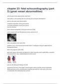
what is this image showing
Apical cardiac imaging including the outflow tracts shows d-transposition of the great arteries
(d-TGA). The pulmonary artery (pa) arises from the left ventricle (LV) and the aorta (ao) from the
right ventricle. At right: Color flow imaging of d-TGA shows that both outflow tracts are virtually
side-by-side and not crossing
what is corrected transposition of the great arteries (I-TGA)
in congenitally corrected transposition the position of the two ventricles is reversed so that the
right atrium connects to the left ventricle and the left atrium connects to the right ventricle
what is connected in I-TGA
5) (great vessel abnormalities)
what do great vessel abnormalities result from
interruptions in the spiraling that occurs during early embryonic development
what are the great vessel abnormalities
complete transposition of the great arteries
corrected transposition of the great arteries
truncus arteriosus
what is dextro-transposition of the great arteries (d-TGA)
aorta connects to RV and the pulmonary artery connects to LV
when are problems seen with d-TGA
problems occur in the neonatal period when there is inadequate mixing of oxygenated and
deoxygenated blood
what is the prognosis with d-TGA
good with surgical intervention but depends on other anomalies (atrial septal defect, anomalies
of atrioventricular valve, underdevelopment of RV and LV)
what are the ultrasound findings of d-TGA
parasternal short axis is key image to view the great arteries and their normal relationship
(RVOT, pulmonary artery, and bifurcation should be seen anterior to the aorta in this view)
in transposition this relationship does not exist
, what is the relationship of vessels in d-TGA
impossible to demonstrate the bifurcation of the PA because the aorta is the anterior vessel
crisscross sweep of the great arteries is not possible
great arteries are sometimes seen parallel to each other as they arise from the ventricles
what is this image showing
Apical cardiac imaging including the outflow tracts shows d-transposition of the great arteries
(d-TGA). The pulmonary artery (pa) arises from the left ventricle (LV) and the aorta (ao) from the
right ventricle. At right: Color flow imaging of d-TGA shows that both outflow tracts are virtually
side-by-side and not crossing
what is corrected transposition of the great arteries (I-TGA)
in congenitally corrected transposition the position of the two ventricles is reversed so that the
right atrium connects to the left ventricle and the left atrium connects to the right ventricle
what is connected in I-TGA