Epitheel
Bestaat uit cellen die goed aaneen sluiten (hebben weinig ECM)
Belangrijk: zo kan niets van ene plaats zomaar naar andere plaats
Bv. Vanuit mondholte naar onderliggende weefsels
Je hebt dunne en dikke epithelen (te maken met hoeveel druk ergens wordt uitgeoefend)
Epitheelcellen : gepolariseerde cellen
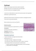
Epitheel ligt op basaal membraan (verankering van epitheel in onderliggende weefsels)
Onder basaal membraan ligt bindweefsel
Basale membraan kan je niet zien op LM (wel EM)
Bedekkend epitheel
Vormt een scheiding tussen compartimenten (o.a. rond alle buizen in lichaam)
Klierepitheel
Secretiefunctie (zweetklier, speekselklier…)
Myoepitheel: spierfunctie
Neuroepitheel: zenuwfunctie
Bedekkend epitheel
= laag die uitwendige en inwendige lichaamsholten bedekt
Wordt beschreven a.d.h.v.:
- aantal lagen (meerlagig – eenlagig-pseudomeerlagig )
- vorm cellen (plat = plaveisel, kubusvormig = kubisch, cilinders = cilindrisch)
- oppervlaktespecialisaties aan de apicale zijde van epitheelweefsel
o Microvilli = plooiing voor opp vergroting (op plaatsen met veel uitwisseling)
o Trilharen/cillien voor voortbeweging
,Éénlagige epithelen
1-lagig plaveiselepitheel
Platte polygonale cellen met apicaal uitpuilende kernen
Voorkomen: plaatsen waar veel uitwisseling gebeurt
Bloedvaten, lichaamsholten, longblaasjes, nierbuisjes, binnenzijde cornea (=hoornvlies)
Endotheel: bloedvaten (speciale functies zoals bloedstolling)
Mesotheel: buikvlies, longvlies, hartvlies (microvilli)
(zorgt voor toelaten beweging bv ademen en uitzetten van hart)
Functie:
Diffusie, resorptie, secretie
, Éénlagig kubisch epitheel
Kubusvorm
Evenveel cytoplasma boven en onder kern (centrale kern)
Plaats:
Klierbuisjes, nierbuisjes (microvilli), retina
Functie:
Resorptie, secretie, lichtregulatie (retina)
Éénalgig cylindrisch epitheel
Hogere cellen
Ovale kern die basaal gelegen is: meer
cytoplasma naar boven
Plaats:
Epitheel van spijsverteringsstelsel (microvilli)
Afvoergangen van klieren, nierbuisjes, eileider
(trilharen)
Functie:
Resorptie, secretie, voortbeweging (eileider)
Bestaat uit cellen die goed aaneen sluiten (hebben weinig ECM)
Belangrijk: zo kan niets van ene plaats zomaar naar andere plaats
Bv. Vanuit mondholte naar onderliggende weefsels
Je hebt dunne en dikke epithelen (te maken met hoeveel druk ergens wordt uitgeoefend)
Epitheelcellen : gepolariseerde cellen
Epitheel ligt op basaal membraan (verankering van epitheel in onderliggende weefsels)
Onder basaal membraan ligt bindweefsel
Basale membraan kan je niet zien op LM (wel EM)
Bedekkend epitheel
Vormt een scheiding tussen compartimenten (o.a. rond alle buizen in lichaam)
Klierepitheel
Secretiefunctie (zweetklier, speekselklier…)
Myoepitheel: spierfunctie
Neuroepitheel: zenuwfunctie
Bedekkend epitheel
= laag die uitwendige en inwendige lichaamsholten bedekt
Wordt beschreven a.d.h.v.:
- aantal lagen (meerlagig – eenlagig-pseudomeerlagig )
- vorm cellen (plat = plaveisel, kubusvormig = kubisch, cilinders = cilindrisch)
- oppervlaktespecialisaties aan de apicale zijde van epitheelweefsel
o Microvilli = plooiing voor opp vergroting (op plaatsen met veel uitwisseling)
o Trilharen/cillien voor voortbeweging
,Éénlagige epithelen
1-lagig plaveiselepitheel
Platte polygonale cellen met apicaal uitpuilende kernen
Voorkomen: plaatsen waar veel uitwisseling gebeurt
Bloedvaten, lichaamsholten, longblaasjes, nierbuisjes, binnenzijde cornea (=hoornvlies)
Endotheel: bloedvaten (speciale functies zoals bloedstolling)
Mesotheel: buikvlies, longvlies, hartvlies (microvilli)
(zorgt voor toelaten beweging bv ademen en uitzetten van hart)
Functie:
Diffusie, resorptie, secretie
, Éénlagig kubisch epitheel
Kubusvorm
Evenveel cytoplasma boven en onder kern (centrale kern)
Plaats:
Klierbuisjes, nierbuisjes (microvilli), retina
Functie:
Resorptie, secretie, lichtregulatie (retina)
Éénalgig cylindrisch epitheel
Hogere cellen
Ovale kern die basaal gelegen is: meer
cytoplasma naar boven
Plaats:
Epitheel van spijsverteringsstelsel (microvilli)
Afvoergangen van klieren, nierbuisjes, eileider
(trilharen)
Functie:
Resorptie, secretie, voortbeweging (eileider)