Infection and Immunity
Immunology
• Physiological mechanisms that defend against invasion by other organisms
• A definition of immunology. However, immunology is much more than this.
Pathogens
• Infectious organisms (or viruses) that cause disease
• Habitual (Typhoid bacillus)
• Opportunistic – when your immune system isn’t working properly (immune deficiencies)
Different classes of pathogens, viruses, bacteria, single celled eukaryotes, multicellular organisms.
• Fungi infections – ring worm. Viral infections – influenza, HIV. Bacterial infections – Staphylococcus
First line of defence - to stop the infectious organism getting into the body. The skin and
mucosa provide well maintained mechanical, chemical and microbiological barriers that
prevent most pathogen s from gaining access to the cells and tissue of the body. When this
barrier is broken and pathogens gain entry to the body’s soft tissues, the fixed defences of
the immune system are triggered.
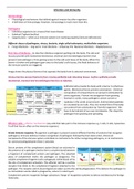
Image shows the physical barriers that separate the body from its external environment.
Various barriers prevent bacteria from crossing epithelial and colonizing tissues. Surface epithelia provide
mechanical, chemical and microbiological barriers to infection.
Epithelial cells provide the body with a barrier to infectious
agents. Mechanical forces prevent colonization. Chemical
composition of compartments can prevent colonization by
some organisms. Prevent microorganism from growing.
Stomach is acidic, many pathogens cannot survive or
replicate in this acidic environment. Antimicrobial peptides
are secreted by our cells. Also, the normal flora of the body
can protect from colonization by ‘harmful organisms’. We
have to be tolerant of our own microbiome. This stops the
pathogen from growing and spreading.
Effector cells – effector mechanisms (any cells that take part in the immune response e.g. t cells, b cells, lysosomes.
A cell that has an effect on the immune response.)
Innate immune response: Recognition a pathogen is present (several different families of proteins that recognise
pathogens). Immune defence involves recognition of pathogens followed by their destruction. Almost all
components of the immune system contribute to mechanisms for either recognizing pathogens, or to mechanisms
for communicating between these 2 activates.
Serum proteins of the complement system (blue) are activated in
the presence of a pathogen (red) to form a covalent bond between
a fragment of complement protein and pathogen. The attached
piece of complement marks the pathogen as dangerous. The
soluble complement fragment calls a phagocytic white blood cell to
the site of complement activation. This effector cell has a surface
receptor that binds to the complement fragment attached to the
pathogen. The receptor and its bound ligand are taken up into the cell by phagocytosis. This delivers the pathogen to
an intracellular vesicle called a phagosome, where it is destroyed.
Complement system recognises foreign substances. Range of recognition systems to detect and destroy the foreign
molecules.
, Inflammation
The innate immune system response causes inflammation at
site of infection.
Innate immune mechanism established a state of
inflammation at sites of infection. Image shows the events
following an abrasion of the skin. Bacteria invade underlying
connective tissue and stimulate the innate immune system.
Inflammation is often due to an immune reaction in response to infection. However, inflammation is not always due
to invasion by a foreign organism. Can get autoimmune (inflammatory) responses. Can get inflammation without
disease e.g. artritus. White blood cells that are usually present in inflamed tissues and release substance that
contribute to inflamation are called inflammatory cells.
Bacteria have a route to get into the tissues of the body via the wound. Once in the tissues, the bloody clotting will
prevent more infection. Effector send out signals (cytokines). Cytokines increase the permeabily of the endothelium
cells that line the blood vessels.
• Surface bound introduces bacteria, which activates resident effect cells to secrete cytokines.
• Vasodilation and increased vascular permeability allow fluid, protein and inflammatory cells to leave
blood and enter tissue
• The infected tissue becomes inflamed, causing redness, heat, swelling and pain.
Septis – immmune reaction. The events occur all over the body (not localised). This could cause
overactivation of the immune systme.
Adaptive immune response adds to ongoing innate response
The adaptive immune response adds to an ongoing innate
immune response.
Innate immune system can respond rapidly. This is a fixed
response, it does not adapt. It recognises something as foreign in
a limited number of ways. Innate immune system doesn’t
recognise individual proteins as foreign.
Adaptive response – directed towards a particular pathogen (very specific). This only occurs once you have been
effected. First time infected = slow response, do no respond immediately. Certain cells get selected, these then have
to replicate and increase in number. Once pathogen has been removed, memory cells remember the pathogen and
next time will clear the pathogens very quickly before you get any symptoms.
Immune responses are not always beneficial - our immune system does not always function ideally. It can attack
our own organs/tissues (autoimmunity) and or respond inappropriately to harmless challenges (allergy). Sometimes
our immune system can get out of control and sepsis can result. Recognising self from non-self.
Cells of the immune system
Immune system cells with different functions all derive from hematopoietic stem cells (HSC).
Adaptive – lymphoid. Recognise by the type of proteins they express on their cell surface. Antibodies will bind
specifically to these proteins.
, The HSC divides and differentiates into more specialised progenitor cells that
give rise to the lymphoid family, the myeloid family and the erythroid
family/lineage.
On activation by infection, B cells divide and differentiate into plasma cells,
whereas T cells differentiate into various type of effector T cells. The myeloid
progenitors cell divides and differentiates to produce at least 6 cell types.
Each CD refer to a different surface protein. Cell types can often be distinguished by
their cell surface (CD) markers. In immunology CD markers (cluster of differentiation)
and combinations of markers are used to define cell types. Unfortunately the
nomenclature is not always intuitive.
Flow Cytometry
Flow cytometry is a technology that is used to analyse the physical and
chemical characteristics of particles in a fluid as it passes through at least one
laser. Cell components are fluorescently labelled and then excited by the laser
to emit light at varying wavelengths.
Flow cytometry is a technique used commonly in immunology to analyze the
constitution of populations of cells. FACS (Fluorescence activated cell sorting)
can separate cells into different populations. Uses CD markers.
Neutrophils are stored in the bone marrow and move in large numbers to
sites of infection, where they act and then die.
Neutrophils are the most abundant of effector cells. They are phagocytes
summoned to sites of infection during an immune response.
After one round of ingestion and killing of bacteria, a neutrophil dies. The dead
neutrophils are eventually mopped up by long loved tissue macrophages,
which break them down. Pus is composed of dead neutrophils.
Macrophages respond to pathogens by using different receptors to stimulate phagocytosis and cytokine secretion.
Macrophages sense infection and alert the immune system. Induce inflammation. Have many different receptors to
recognize ‘foreign’ organisms. The left image shows receptor mediated phagocytosis of bacteria by a macrophage.
The bacterium (red) binds to cell surface receptors (blue) on the
macrophage, inducing engulfment of the bacterium into a phagosome
within the macrophage cytoplasm. Fusion of the phagosome with
lysosomes forms and acidic vesicle (phagolysosome), which contains toxic
small molecules and hydrolytic enzymes that kill and degrade the
bacterium. The right image shows how a bacterial component binding to a
different type of cell surface receptors sends a signal to the macrophage’s
nucleus that initiates the transcription of genes for inflammatory
cytokines. Cytokines are then secreted into the extracellular matrix.
, The relative abundance of the leukocyte cell types in human peripheral blood. Typical
proportions of circulating immune cells.
Most lymphocytes are present in specialised lymphoid tissues
• Primary lymphoid tissues:- Where lymphocytes develop to a stage at which they are able to respond to a
pathogen.
• Secondary lymphoid tissues:- where mature lymphocytes are stimulated to
respond to pathogen
Sites of principal lymphoid tissues
Small lymphocytes are unique amongst blood cells. Found in lymph vessels as well as
blood. Lymphocytes arise from stem cells in the bone marrow. B cells complete their
maturation in the bone marrow, whereas T cells leave at an immature stage and
complete their development in the thymus. The bone marrow and the thymus are the
primary lymphoid tissues (red). Secondary lymphoid tissues are yellow and black lines
are the lymphatics.
Lymphocyte recirculation
Small lymphocytes are unique among blood cells in travelling through the body in the
lymph as well as the blood. Lymphocytes leave the blood through the walls of fine
capillaries in secondary lymphoid organs. After spending some time in the lymph
node, lymphocytes leave in the efferent lymph and return to the blood at the left
subclavian vein. If a lymphocyte in a lymph node encounters a pathogen to which its
cell-surface receptor binds to, it stops recirculating.
Innate and Adaptive immunity
• Adaptive immunity is initiated in secondary lymphoid tissues. Innate and adaptive
immunity work together to defend the body.
• Circulating lymphocytes meet lymph-borne pathogens in draining lymph nodes.
Image - Activation of adaptive immunity in the drainage node.
• Pathogens, pathogen components, and dendritic cells carrying pathogens and molecules
derived from them arrive in the afferent lymph draining the site of infection.
• Free pathogens and debris are removed by macrophages. The dendritic cells become
resident in the lymph node and move to the T cell areas, where they meet small
lymphocytes that have entered the node from the blood (green).
• The dendritic cells specifically stimulate the division and differentiation of pathogen-
specific small lymphocytes into effector lymphocytes (blue).
• Some helper T cells and cytotoxic T cells leave in the efferent lymph and travel to the
infected tissue via the lymph and the blood. Other helper T cells remain in the lymph node
and stimulate the division and differentiation of pathogen-specific B cells into plasma cells
(yellow).
• Plasma cells move to the medulla of the lymph node, where they secrete pathogen-
specific antibodies, which are taken to the site of infection by the efferent lymph and then
to the blood. Some plasma cells leave the lymph node and travel via efferent lymph and
the blood to the bone marrow, where they continue to secrete antibodies.
This is a a possible scenario in the clearance of an infection.
Immunology
• Physiological mechanisms that defend against invasion by other organisms
• A definition of immunology. However, immunology is much more than this.
Pathogens
• Infectious organisms (or viruses) that cause disease
• Habitual (Typhoid bacillus)
• Opportunistic – when your immune system isn’t working properly (immune deficiencies)
Different classes of pathogens, viruses, bacteria, single celled eukaryotes, multicellular organisms.
• Fungi infections – ring worm. Viral infections – influenza, HIV. Bacterial infections – Staphylococcus
First line of defence - to stop the infectious organism getting into the body. The skin and
mucosa provide well maintained mechanical, chemical and microbiological barriers that
prevent most pathogen s from gaining access to the cells and tissue of the body. When this
barrier is broken and pathogens gain entry to the body’s soft tissues, the fixed defences of
the immune system are triggered.
Image shows the physical barriers that separate the body from its external environment.
Various barriers prevent bacteria from crossing epithelial and colonizing tissues. Surface epithelia provide
mechanical, chemical and microbiological barriers to infection.
Epithelial cells provide the body with a barrier to infectious
agents. Mechanical forces prevent colonization. Chemical
composition of compartments can prevent colonization by
some organisms. Prevent microorganism from growing.
Stomach is acidic, many pathogens cannot survive or
replicate in this acidic environment. Antimicrobial peptides
are secreted by our cells. Also, the normal flora of the body
can protect from colonization by ‘harmful organisms’. We
have to be tolerant of our own microbiome. This stops the
pathogen from growing and spreading.
Effector cells – effector mechanisms (any cells that take part in the immune response e.g. t cells, b cells, lysosomes.
A cell that has an effect on the immune response.)
Innate immune response: Recognition a pathogen is present (several different families of proteins that recognise
pathogens). Immune defence involves recognition of pathogens followed by their destruction. Almost all
components of the immune system contribute to mechanisms for either recognizing pathogens, or to mechanisms
for communicating between these 2 activates.
Serum proteins of the complement system (blue) are activated in
the presence of a pathogen (red) to form a covalent bond between
a fragment of complement protein and pathogen. The attached
piece of complement marks the pathogen as dangerous. The
soluble complement fragment calls a phagocytic white blood cell to
the site of complement activation. This effector cell has a surface
receptor that binds to the complement fragment attached to the
pathogen. The receptor and its bound ligand are taken up into the cell by phagocytosis. This delivers the pathogen to
an intracellular vesicle called a phagosome, where it is destroyed.
Complement system recognises foreign substances. Range of recognition systems to detect and destroy the foreign
molecules.
, Inflammation
The innate immune system response causes inflammation at
site of infection.
Innate immune mechanism established a state of
inflammation at sites of infection. Image shows the events
following an abrasion of the skin. Bacteria invade underlying
connective tissue and stimulate the innate immune system.
Inflammation is often due to an immune reaction in response to infection. However, inflammation is not always due
to invasion by a foreign organism. Can get autoimmune (inflammatory) responses. Can get inflammation without
disease e.g. artritus. White blood cells that are usually present in inflamed tissues and release substance that
contribute to inflamation are called inflammatory cells.
Bacteria have a route to get into the tissues of the body via the wound. Once in the tissues, the bloody clotting will
prevent more infection. Effector send out signals (cytokines). Cytokines increase the permeabily of the endothelium
cells that line the blood vessels.
• Surface bound introduces bacteria, which activates resident effect cells to secrete cytokines.
• Vasodilation and increased vascular permeability allow fluid, protein and inflammatory cells to leave
blood and enter tissue
• The infected tissue becomes inflamed, causing redness, heat, swelling and pain.
Septis – immmune reaction. The events occur all over the body (not localised). This could cause
overactivation of the immune systme.
Adaptive immune response adds to ongoing innate response
The adaptive immune response adds to an ongoing innate
immune response.
Innate immune system can respond rapidly. This is a fixed
response, it does not adapt. It recognises something as foreign in
a limited number of ways. Innate immune system doesn’t
recognise individual proteins as foreign.
Adaptive response – directed towards a particular pathogen (very specific). This only occurs once you have been
effected. First time infected = slow response, do no respond immediately. Certain cells get selected, these then have
to replicate and increase in number. Once pathogen has been removed, memory cells remember the pathogen and
next time will clear the pathogens very quickly before you get any symptoms.
Immune responses are not always beneficial - our immune system does not always function ideally. It can attack
our own organs/tissues (autoimmunity) and or respond inappropriately to harmless challenges (allergy). Sometimes
our immune system can get out of control and sepsis can result. Recognising self from non-self.
Cells of the immune system
Immune system cells with different functions all derive from hematopoietic stem cells (HSC).
Adaptive – lymphoid. Recognise by the type of proteins they express on their cell surface. Antibodies will bind
specifically to these proteins.
, The HSC divides and differentiates into more specialised progenitor cells that
give rise to the lymphoid family, the myeloid family and the erythroid
family/lineage.
On activation by infection, B cells divide and differentiate into plasma cells,
whereas T cells differentiate into various type of effector T cells. The myeloid
progenitors cell divides and differentiates to produce at least 6 cell types.
Each CD refer to a different surface protein. Cell types can often be distinguished by
their cell surface (CD) markers. In immunology CD markers (cluster of differentiation)
and combinations of markers are used to define cell types. Unfortunately the
nomenclature is not always intuitive.
Flow Cytometry
Flow cytometry is a technology that is used to analyse the physical and
chemical characteristics of particles in a fluid as it passes through at least one
laser. Cell components are fluorescently labelled and then excited by the laser
to emit light at varying wavelengths.
Flow cytometry is a technique used commonly in immunology to analyze the
constitution of populations of cells. FACS (Fluorescence activated cell sorting)
can separate cells into different populations. Uses CD markers.
Neutrophils are stored in the bone marrow and move in large numbers to
sites of infection, where they act and then die.
Neutrophils are the most abundant of effector cells. They are phagocytes
summoned to sites of infection during an immune response.
After one round of ingestion and killing of bacteria, a neutrophil dies. The dead
neutrophils are eventually mopped up by long loved tissue macrophages,
which break them down. Pus is composed of dead neutrophils.
Macrophages respond to pathogens by using different receptors to stimulate phagocytosis and cytokine secretion.
Macrophages sense infection and alert the immune system. Induce inflammation. Have many different receptors to
recognize ‘foreign’ organisms. The left image shows receptor mediated phagocytosis of bacteria by a macrophage.
The bacterium (red) binds to cell surface receptors (blue) on the
macrophage, inducing engulfment of the bacterium into a phagosome
within the macrophage cytoplasm. Fusion of the phagosome with
lysosomes forms and acidic vesicle (phagolysosome), which contains toxic
small molecules and hydrolytic enzymes that kill and degrade the
bacterium. The right image shows how a bacterial component binding to a
different type of cell surface receptors sends a signal to the macrophage’s
nucleus that initiates the transcription of genes for inflammatory
cytokines. Cytokines are then secreted into the extracellular matrix.
, The relative abundance of the leukocyte cell types in human peripheral blood. Typical
proportions of circulating immune cells.
Most lymphocytes are present in specialised lymphoid tissues
• Primary lymphoid tissues:- Where lymphocytes develop to a stage at which they are able to respond to a
pathogen.
• Secondary lymphoid tissues:- where mature lymphocytes are stimulated to
respond to pathogen
Sites of principal lymphoid tissues
Small lymphocytes are unique amongst blood cells. Found in lymph vessels as well as
blood. Lymphocytes arise from stem cells in the bone marrow. B cells complete their
maturation in the bone marrow, whereas T cells leave at an immature stage and
complete their development in the thymus. The bone marrow and the thymus are the
primary lymphoid tissues (red). Secondary lymphoid tissues are yellow and black lines
are the lymphatics.
Lymphocyte recirculation
Small lymphocytes are unique among blood cells in travelling through the body in the
lymph as well as the blood. Lymphocytes leave the blood through the walls of fine
capillaries in secondary lymphoid organs. After spending some time in the lymph
node, lymphocytes leave in the efferent lymph and return to the blood at the left
subclavian vein. If a lymphocyte in a lymph node encounters a pathogen to which its
cell-surface receptor binds to, it stops recirculating.
Innate and Adaptive immunity
• Adaptive immunity is initiated in secondary lymphoid tissues. Innate and adaptive
immunity work together to defend the body.
• Circulating lymphocytes meet lymph-borne pathogens in draining lymph nodes.
Image - Activation of adaptive immunity in the drainage node.
• Pathogens, pathogen components, and dendritic cells carrying pathogens and molecules
derived from them arrive in the afferent lymph draining the site of infection.
• Free pathogens and debris are removed by macrophages. The dendritic cells become
resident in the lymph node and move to the T cell areas, where they meet small
lymphocytes that have entered the node from the blood (green).
• The dendritic cells specifically stimulate the division and differentiation of pathogen-
specific small lymphocytes into effector lymphocytes (blue).
• Some helper T cells and cytotoxic T cells leave in the efferent lymph and travel to the
infected tissue via the lymph and the blood. Other helper T cells remain in the lymph node
and stimulate the division and differentiation of pathogen-specific B cells into plasma cells
(yellow).
• Plasma cells move to the medulla of the lymph node, where they secrete pathogen-
specific antibodies, which are taken to the site of infection by the efferent lymph and then
to the blood. Some plasma cells leave the lymph node and travel via efferent lymph and
the blood to the bone marrow, where they continue to secrete antibodies.
This is a a possible scenario in the clearance of an infection.