

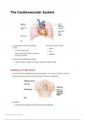
The Cardiovascular System
a closed system of the heart and blood three types of blood vessels
vessels
arteries
the heart pumps blood
veins
blood vessels allow blood to circulate to
capillaries
all parts of the body
functions of the cardiovascular system
transport oxygen, nutrients, cell wastes, hormones to and from cells
anatomy of the heart
size of a human fist, weighing less than a pound (approx. 12 cm long, 8 cm wide, 6 cm thick)
located in the thoracic cavity, between the lungs in the inferior mediastinum
orientation
apex is directed toward the left hip and rests on the diaphragm
The Cardiovascular System 1
, apex location: left of the sternum between the junction of the 4th and 5th ribs near the
articulation of the coastal cartilage
base points toward the right shoulder
location: located at the level of the third coastal cartilage
important to know this when placing the stethoscope
coverings of the heart walls of the heart
pericardial cavity - serous fluid fills the space epicardium
between the layers of the pericardium;
outside layer; the visceral pericardium
important to lubricate the heart, prevent
friction between heart and surrounding thin layer of serous membrane that helps
organs, hold heart in position and maintain to lubricate and protect the outside of
hollow space to expand when full heart
pericardium - a double-walled sac myocardium
fibrous pericardium is loose and middle layer
superficial mostly cardiac muscle
serous membrane is deep to the fibrous makes up majority of mass and thickness
pericardium and composed of two layers of heart wall
1. parietal pericardium - outside layer that responsible for pumping blood
lines the inner surface of the fibrous
endocardium
pericardium
inner layer known as endothelium (simple
2. visceral pericardium - next to heart; aka
squamous)
epicardium
smooth and responsible for keeping blood
from sticking to the inside of the heart and
forming potential deadly blood clots
chambers and associated great vessels
The Cardiovascular System 2
, 4 chambers of the heart heart functions as a double pump
atria (right and left) arteries carry blood away from the heart /
toward the organs
receiving chambers
veins carry blood toward the heart
assist with filling the ventricles with blood
double pump
blood enters under low pressure
right side works as a pulmonary circuit
ventricles (right and left)
pump
discharging chamber
left side works as the systemic circuit
thick-walled pumps of the heart pump
during contraction, blood is propelled into
circulation
septum
The Cardiovascular System 3
a closed system of the heart and blood three types of blood vessels
vessels
arteries
the heart pumps blood
veins
blood vessels allow blood to circulate to
capillaries
all parts of the body
functions of the cardiovascular system
transport oxygen, nutrients, cell wastes, hormones to and from cells
anatomy of the heart
size of a human fist, weighing less than a pound (approx. 12 cm long, 8 cm wide, 6 cm thick)
located in the thoracic cavity, between the lungs in the inferior mediastinum
orientation
apex is directed toward the left hip and rests on the diaphragm
The Cardiovascular System 1
, apex location: left of the sternum between the junction of the 4th and 5th ribs near the
articulation of the coastal cartilage
base points toward the right shoulder
location: located at the level of the third coastal cartilage
important to know this when placing the stethoscope
coverings of the heart walls of the heart
pericardial cavity - serous fluid fills the space epicardium
between the layers of the pericardium;
outside layer; the visceral pericardium
important to lubricate the heart, prevent
friction between heart and surrounding thin layer of serous membrane that helps
organs, hold heart in position and maintain to lubricate and protect the outside of
hollow space to expand when full heart
pericardium - a double-walled sac myocardium
fibrous pericardium is loose and middle layer
superficial mostly cardiac muscle
serous membrane is deep to the fibrous makes up majority of mass and thickness
pericardium and composed of two layers of heart wall
1. parietal pericardium - outside layer that responsible for pumping blood
lines the inner surface of the fibrous
endocardium
pericardium
inner layer known as endothelium (simple
2. visceral pericardium - next to heart; aka
squamous)
epicardium
smooth and responsible for keeping blood
from sticking to the inside of the heart and
forming potential deadly blood clots
chambers and associated great vessels
The Cardiovascular System 2
, 4 chambers of the heart heart functions as a double pump
atria (right and left) arteries carry blood away from the heart /
toward the organs
receiving chambers
veins carry blood toward the heart
assist with filling the ventricles with blood
double pump
blood enters under low pressure
right side works as a pulmonary circuit
ventricles (right and left)
pump
discharging chamber
left side works as the systemic circuit
thick-walled pumps of the heart pump
during contraction, blood is propelled into
circulation
septum
The Cardiovascular System 3


