Lethal Rhythms
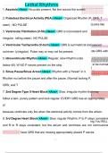
1. Asystole <Ans> No pulse present, flat line across the screen.
2. Pulseless Electrical Activity (PEA) <Ans> Organized Rhythm (P, QRS, T
seen) - NO PULSE (Looks like
3. Ventricular Fibrillation (V-fib) <Ans> QRS is inconsistent and
irregular. rolling water). NO PULSE
4. Ventricular Tachycardia (V-tach) <Ans> QRS is symmetrical and spaced
out even- ly(regular). Pulse may or may not be present. ide QRS with
5. Idioventricular Rhythm <Ans> Regular, slow Rhythm (rate
below 40). W NO P waves present on the strip. st be normal
)
6. Sinus Pause/Sinus Arrest <Ans> Rhythm with a "break" in it.
Rhythm mu before the pause and after the pause. (Normal looking P,
QRS, and T
7. 2nd Degree Type II Heart Block <Ans> Slow, irregular rhythm that may
aced P wave,
follow a tem- porary pattern and look regular. EVERY QRS has an appropriately
pl
because ventricles only fire when the electrical activity comes from the atrium.
8. 3rd Degree Heart Block <Ans> Slow, regular Rhythm. P to P stays consistent
ing.You WILL
and R to R stays consistent, but the atrium and ventricles are not communicat
have QRS that are missing appropriately placed P waves.
1/1
1. Asystole <Ans> No pulse present, flat line across the screen.
2. Pulseless Electrical Activity (PEA) <Ans> Organized Rhythm (P, QRS, T
seen) - NO PULSE (Looks like
3. Ventricular Fibrillation (V-fib) <Ans> QRS is inconsistent and
irregular. rolling water). NO PULSE
4. Ventricular Tachycardia (V-tach) <Ans> QRS is symmetrical and spaced
out even- ly(regular). Pulse may or may not be present. ide QRS with
5. Idioventricular Rhythm <Ans> Regular, slow Rhythm (rate
below 40). W NO P waves present on the strip. st be normal
)
6. Sinus Pause/Sinus Arrest <Ans> Rhythm with a "break" in it.
Rhythm mu before the pause and after the pause. (Normal looking P,
QRS, and T
7. 2nd Degree Type II Heart Block <Ans> Slow, irregular rhythm that may
aced P wave,
follow a tem- porary pattern and look regular. EVERY QRS has an appropriately
pl
because ventricles only fire when the electrical activity comes from the atrium.
8. 3rd Degree Heart Block <Ans> Slow, regular Rhythm. P to P stays consistent
ing.You WILL
and R to R stays consistent, but the atrium and ventricles are not communicat
have QRS that are missing appropriately placed P waves.
1/1