Neuropsychology & psychopharmacology – januari 2025 1
PSYCHOPHARMACOLOGY
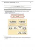
STRUCTURE AND FUNCTION OF THE NERVOUS SYSTEM
The nervous system is responsible for the reception and processing of sensory information from both external
and internal environments, divided into two major parts:
- Central nervous system (CNS): the brain, protected by the skull, directly connected to the spinal cord,
protected by a vertebral column or spine
- Peripheral nervous system (PNS): cranial and spinal nerves
This division is arbitrary because the systems work together and are connected
The nervous system has 3 specific functions:
1. Receiving sensory input
2. Processing and integration of information by summation, reviewing, saving and creating appropriate motor
responses
3. Generating motor output
,Neuropsychology & psychopharmacology – januari 2025 2
NERVOUS TISSUE AND ANATOMY
Nervous tissue contains two types of cells
NEURONS
= cells that transmit nerve impulses between parts of the nervous
system
- Sensory neurons: takes nerve impulses all the way from
sensory receptors to the CNS
- Motor neurons: takes nerve impulses from the CNS to an
effector (like an organ, muscle or gland)
- Interneurons: takes input from sensory neurons or other
interneurons to motor neurons and so lies entirely in the CNS
• Can sometimes have a short axon that is not covered in
myelin sheath
Neurons vary in appearance, but all have
- Cell body: contains the nucleus as well as other organelles
- Dendrites: short extensions that receive signals from sensory
receptors or other neurons
- Axon or nerve fiber: conducts nerve impulses resulting from
the received signals in dendrites
• Collectively nerve fibers form a nerve
• Is usually long and covered by a white-looking myelin
sheath, though some interneurons have a short axon that
is not covered
• Myelin sheath plays an important role in the rate at which
signals move through the neuron and for nerve
regeneration within the PNS
NEUROGLIA OR GLIAL CELLS
= cells that support and nourish neurons (and so there are many more)
• Microglia: phagocytic cells that help remove bacteria and debris
• Astrocytes: provide metabolic and structural support directly to the neurons
• Schwann cells: form the myelin sheath and nodes of Ranvier in the PNS
• Oligodendrocytes: form the myelin sheath and nodes of Ranvier in the CNS
NEURON PHYSIOLOGY
RESTING POTENTIAL
= the potential energy that a resting neuron (that is not conducting a nerve
impulse) has because the plasma membrane of the neuron is polarized
- The outside of the cell is positively charged because positively charged
sodium ions (Na+) are clustered around it, while the inside is negatively
charged because of large, negatively charged proteins and other
molecules trapped inside because of their size
• In its resting state, the plasma membrane is permeable to potassium
(K+) but not to sodium, causing the potassium to contribute to the
positive charge by diffusing out of the cell to join the sodium ions
- The difference in electrical charge between the inside and outside of the
cell is the resting membrane potential, usually around -70 millivolts (mV)
or -0,070 volts (V)
,Neuropsychology & psychopharmacology – januari 2025 3
• Neurons must maintain their resting potential to be able to work and do so by transporting sodium
ions out of the cell and importing potassium ions via a sodium-potassium pump, which recharges the
cell
ACTION POTENTIAL
= a rapid and transient change in the electrical potential across a neuron's
membrane, which allows the neuron to transmit information over long
distances
1. Action potential is triggered when a neuron receives a stimulus strong
enough to depolarize the membrane, with a threshold of -55 mV
• If the threshold is not reached, action potential does not occur
• Increasing the strength of a stimulus (e.g. squeezing someone’s arm
harder) does not change the strength of an action potential but could
cause more action potentials to occur and therefore change
someone’s perception (e.g. experiencing more pain)
2. Depolarization will occur when the protein channels open and sodium
ions rush into the cell, reaching around +35 mV
3. Repolarization will start almost immediately after when the sodium
channels close and the potassium protein channels open, causing the potassium to flow out of the cell
• The inside of the cell will start to become negative again because of the large, negatively charged ions
trapped inside the cell
4. The sodium-potassium pump completes the action potential by returning the potassium ions to the inside
of the cell and sodium ions to the outside, restoring the resting potential
This entire process requires only 3 to 4 milliseconds to complete
PROPAGATION OF AN ACTION POTENTIAL
= the action potential is self-propagating, meaning it travels down the axon in a chain reaction with each action
potential generating another along the entire length of the axon
- In unmyelinated axons, the depolarization spreads continuously along the membrane but rather slow at ±1
m/s because each section of the axon must be stimulated
- In myelinated axons, the action potential “jumps” between the nodes of Ranvier, speeding up the
conduction to sometimes 100 m/s – this is called salutatory conduction
The intensity of a message is determined by how many action potentials are generated within a given time
As soon as the action potential has passed, that portion of the axon undergoes a short refractory period during
which it is unable to conduct an action potential
This ensures the one-way direction of a signal
SYNAPTIC TRANSMISSION
The synapse is the region where the axon terminal (tips of axon branches) lies close to either the dendrite or cell
body of another neuron
- The synaptic cleft separates the sending neuron from the receiving neuron
- Transmission in the synapse is carried out by molecules called neurotransmitters, stored in synaptic vesicles
in the axon terminals
1) Nerve signals travel along the axon to reach the axon terminal
2) Calcium ions enter the terminal (because of depolarization)
3) Triggered by calcium, the synaptic vesicles fuse with the sending/presynaptic membrane
4) Neurotransmitter molecules release into the synaptic cleft and diffuse across the cleft to the
receiving/postsynaptic membrane, where they bind with specific receptor proteins
5) Synaptic integration: summing up of all the excitatory and inhibitory signals the neuron received
leading to the response of the receiving neuron, which can be toward excitation or toward inhibition
, Neuropsychology & psychopharmacology – januari 2025 4
▪ Excitation occurs because the neurotransmitters have caused sodium ions to diffuse into the
receiving neuron through a now open sodium gate (action potential can occur)
▪ Inhibition occurs if the neurotransmitters have caused potassium ions to exit the receiving neuron
6) Neurotransmitters are broken down by enzymes
7) Reuptake into the presynaptic neuron
8) Diffusion or uptake by other cells
RECEPTORS
1. Ligand-gated ion channels (also known as ionotropic receptors): receptors that bind the transmitter and are
directly coupled to an ion channel, so when the transmitter binds it can immediately cause influx or efflux
of ions, immediately influencing the excitability of the postsynaptic cell
• This is a fast and brief process, meaning they are not useful for the transmitting of information for a
longer period of time
PSYCHOPHARMACOLOGY
STRUCTURE AND FUNCTION OF THE NERVOUS SYSTEM
The nervous system is responsible for the reception and processing of sensory information from both external
and internal environments, divided into two major parts:
- Central nervous system (CNS): the brain, protected by the skull, directly connected to the spinal cord,
protected by a vertebral column or spine
- Peripheral nervous system (PNS): cranial and spinal nerves
This division is arbitrary because the systems work together and are connected
The nervous system has 3 specific functions:
1. Receiving sensory input
2. Processing and integration of information by summation, reviewing, saving and creating appropriate motor
responses
3. Generating motor output
,Neuropsychology & psychopharmacology – januari 2025 2
NERVOUS TISSUE AND ANATOMY
Nervous tissue contains two types of cells
NEURONS
= cells that transmit nerve impulses between parts of the nervous
system
- Sensory neurons: takes nerve impulses all the way from
sensory receptors to the CNS
- Motor neurons: takes nerve impulses from the CNS to an
effector (like an organ, muscle or gland)
- Interneurons: takes input from sensory neurons or other
interneurons to motor neurons and so lies entirely in the CNS
• Can sometimes have a short axon that is not covered in
myelin sheath
Neurons vary in appearance, but all have
- Cell body: contains the nucleus as well as other organelles
- Dendrites: short extensions that receive signals from sensory
receptors or other neurons
- Axon or nerve fiber: conducts nerve impulses resulting from
the received signals in dendrites
• Collectively nerve fibers form a nerve
• Is usually long and covered by a white-looking myelin
sheath, though some interneurons have a short axon that
is not covered
• Myelin sheath plays an important role in the rate at which
signals move through the neuron and for nerve
regeneration within the PNS
NEUROGLIA OR GLIAL CELLS
= cells that support and nourish neurons (and so there are many more)
• Microglia: phagocytic cells that help remove bacteria and debris
• Astrocytes: provide metabolic and structural support directly to the neurons
• Schwann cells: form the myelin sheath and nodes of Ranvier in the PNS
• Oligodendrocytes: form the myelin sheath and nodes of Ranvier in the CNS
NEURON PHYSIOLOGY
RESTING POTENTIAL
= the potential energy that a resting neuron (that is not conducting a nerve
impulse) has because the plasma membrane of the neuron is polarized
- The outside of the cell is positively charged because positively charged
sodium ions (Na+) are clustered around it, while the inside is negatively
charged because of large, negatively charged proteins and other
molecules trapped inside because of their size
• In its resting state, the plasma membrane is permeable to potassium
(K+) but not to sodium, causing the potassium to contribute to the
positive charge by diffusing out of the cell to join the sodium ions
- The difference in electrical charge between the inside and outside of the
cell is the resting membrane potential, usually around -70 millivolts (mV)
or -0,070 volts (V)
,Neuropsychology & psychopharmacology – januari 2025 3
• Neurons must maintain their resting potential to be able to work and do so by transporting sodium
ions out of the cell and importing potassium ions via a sodium-potassium pump, which recharges the
cell
ACTION POTENTIAL
= a rapid and transient change in the electrical potential across a neuron's
membrane, which allows the neuron to transmit information over long
distances
1. Action potential is triggered when a neuron receives a stimulus strong
enough to depolarize the membrane, with a threshold of -55 mV
• If the threshold is not reached, action potential does not occur
• Increasing the strength of a stimulus (e.g. squeezing someone’s arm
harder) does not change the strength of an action potential but could
cause more action potentials to occur and therefore change
someone’s perception (e.g. experiencing more pain)
2. Depolarization will occur when the protein channels open and sodium
ions rush into the cell, reaching around +35 mV
3. Repolarization will start almost immediately after when the sodium
channels close and the potassium protein channels open, causing the potassium to flow out of the cell
• The inside of the cell will start to become negative again because of the large, negatively charged ions
trapped inside the cell
4. The sodium-potassium pump completes the action potential by returning the potassium ions to the inside
of the cell and sodium ions to the outside, restoring the resting potential
This entire process requires only 3 to 4 milliseconds to complete
PROPAGATION OF AN ACTION POTENTIAL
= the action potential is self-propagating, meaning it travels down the axon in a chain reaction with each action
potential generating another along the entire length of the axon
- In unmyelinated axons, the depolarization spreads continuously along the membrane but rather slow at ±1
m/s because each section of the axon must be stimulated
- In myelinated axons, the action potential “jumps” between the nodes of Ranvier, speeding up the
conduction to sometimes 100 m/s – this is called salutatory conduction
The intensity of a message is determined by how many action potentials are generated within a given time
As soon as the action potential has passed, that portion of the axon undergoes a short refractory period during
which it is unable to conduct an action potential
This ensures the one-way direction of a signal
SYNAPTIC TRANSMISSION
The synapse is the region where the axon terminal (tips of axon branches) lies close to either the dendrite or cell
body of another neuron
- The synaptic cleft separates the sending neuron from the receiving neuron
- Transmission in the synapse is carried out by molecules called neurotransmitters, stored in synaptic vesicles
in the axon terminals
1) Nerve signals travel along the axon to reach the axon terminal
2) Calcium ions enter the terminal (because of depolarization)
3) Triggered by calcium, the synaptic vesicles fuse with the sending/presynaptic membrane
4) Neurotransmitter molecules release into the synaptic cleft and diffuse across the cleft to the
receiving/postsynaptic membrane, where they bind with specific receptor proteins
5) Synaptic integration: summing up of all the excitatory and inhibitory signals the neuron received
leading to the response of the receiving neuron, which can be toward excitation or toward inhibition
, Neuropsychology & psychopharmacology – januari 2025 4
▪ Excitation occurs because the neurotransmitters have caused sodium ions to diffuse into the
receiving neuron through a now open sodium gate (action potential can occur)
▪ Inhibition occurs if the neurotransmitters have caused potassium ions to exit the receiving neuron
6) Neurotransmitters are broken down by enzymes
7) Reuptake into the presynaptic neuron
8) Diffusion or uptake by other cells
RECEPTORS
1. Ligand-gated ion channels (also known as ionotropic receptors): receptors that bind the transmitter and are
directly coupled to an ion channel, so when the transmitter binds it can immediately cause influx or efflux
of ions, immediately influencing the excitability of the postsynaptic cell
• This is a fast and brief process, meaning they are not useful for the transmitting of information for a
longer period of time