,Reproductive System
• Its activities affect other systems
28-1 Male and female reproductive system structures produce gametes that
combine
• Continue human existence—by producing, storing, nourishing, and
transporting functional male and female reproductive cells aka gametes
• The reproductive system includes the following basic structures: Gonads,
reproductive organs that produce gametes and hormones, Ducts that receive
and transport the gametes, Accessory glands and organs that secrete fluids
into the reproductive system ducts or into other excretory ducts, Perineal
structures aka external genitalia
• In both males and females, the ducts are connected to chambers and
passageways that open to the outside.
• male and female reproductive systems are functionally different
testicles
• the testes or testicles are male gonads that secrete sex hormones called
androgens. The main androgen is testosterone
• The testes also produce the male gametes = sperm. ~½ billion a day
• During emission, mature sperm travel along a lengthy duct system, where they
are mixed with the secretions of accessory glands.=> creates semen
• During ejaculation, semen is expelled from the body.
• The testes hang within the scrotum, a fleshy pouch suspended inferior to the
perineum.
• The scrotum is anterior to the anus, and posterior to the base of the penis
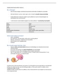
ovaries
• the ovaries = female gonads
• each month it release only one immature gamete, called an oocyte which
travels along one of two short uterine tubes into the uterus (a muscular organ)
,• The ovaries also secrete female sex hormones, including estrogens.
• A short passageway, the vagina connects the uterus with the exterior.
• During intercourse, ejaculation introduces semen into the vagina, and the
sperm then ascend the female reproductive tract. If a sperm reaches the
oocyte and starts the process of fertilization, the oocyte matures into an ovum
(plural, ova). The uterus will then enclose and support the developing embryo
as it grows into a fetus and prepares for birth
28-2 The structures of the male reproductive system = testes + duct system +
accessory glands + penis
• Figure 28–1 shows the main structures of the male reproductive system.
• Starting from a testis, the sperm travel within the male reproductive duct
system, which consists of the epididymis, the ductus deferens, and the urethra
before leaving the body.
• Accessory glands: the seminal glands, the prostate, and the bulbo-urethral
gland
• male external genitalia consist of the scrotum that encloses the testes, the
urethra, and the penis (an erectile organ)
The Spermatic Cords
• The spermatic cords are paired structures extending between the
abdominopelvic cavity and the testes
• Each spermatic cord begins at the entrance to the inguinal canal. After passing
through the inguinal canal, the spermatic cord descends into the scrotum
• 1 spermatic cord consists of layers of fascia and muscle enclosing the ductus
deferens and the blood vessels, nerves, and lymphatic vessels that supply the
testes.
• The blood vessels include the deferential artery, a testicular artery, and the
pampiniform plexus of a testicular vein.
• Branches of the genitofemoral nerve from the lumbar plexus provide
innervation.
• The inguinal canals form during development as the testes descend into the
, scrotum. At that time, these canals link the scrotal cavities with the peritoneal
cavity. In normal adult males, the inguinal canals are closed, but the spermatic
cords create weak points in the abdominal wall that remain throughout life. As
a result, inguinal hernias—protrusions of visceral tissues or organs into the
inguinal canal—are fairly common in males.
The Scrotum and the Position of the Testes
• The scrotum is divided internally into two chambers.
• A raised thickening in the scrotal surface known as the raphe of scrotum
divides it in two (see Figure 28–2).
• Each testis lies in a separate chamber, or scrotal cavity. cavities are separated
by a partition,
• A narrow space separates the inner surface of the scrotum from the outer
surface of the testis.
• The tunica vaginalis a serous membrane, lines the scrotal cavity and reduces
friction between the opposing parietal (outer) layer and visceral (inner) layer
• scrotum = thin layer of skin + the underlying superficial fascia.
• The dermis contains a layer of smooth muscle, the dartos muscle.
• dartos muscle = Resting muscle tone elevates the testes and causes wrinkling
• Cremaster (skeletal) muscle lies deep to the dermis. cremasteric reflex
• cremaster contracts tenses and pulls the testes closer to the body; when:
arousal, decreased temperature stroke the skin on the upper thigh
• Sperm development testes must be about 1.1°C (2°F) lower than the body
Gross Anatomy of the Testes
• Deep to the tunica vaginalis covering the testis is the tunica albuginea a dense
layer of CT rich in collagen fibers
• These fibers are continuous with those surrounding the adjacent epididymis
and extend into the testis. There they form
• fibrous partitions = septa testis - columns between testis lobes, they converge
toward the entrance to the epididymis. The CTs in this region support the blood
vessels and lymphatic vessels that supply and drain the testis, and the efferent
• Its activities affect other systems
28-1 Male and female reproductive system structures produce gametes that
combine
• Continue human existence—by producing, storing, nourishing, and
transporting functional male and female reproductive cells aka gametes
• The reproductive system includes the following basic structures: Gonads,
reproductive organs that produce gametes and hormones, Ducts that receive
and transport the gametes, Accessory glands and organs that secrete fluids
into the reproductive system ducts or into other excretory ducts, Perineal
structures aka external genitalia
• In both males and females, the ducts are connected to chambers and
passageways that open to the outside.
• male and female reproductive systems are functionally different
testicles
• the testes or testicles are male gonads that secrete sex hormones called
androgens. The main androgen is testosterone
• The testes also produce the male gametes = sperm. ~½ billion a day
• During emission, mature sperm travel along a lengthy duct system, where they
are mixed with the secretions of accessory glands.=> creates semen
• During ejaculation, semen is expelled from the body.
• The testes hang within the scrotum, a fleshy pouch suspended inferior to the
perineum.
• The scrotum is anterior to the anus, and posterior to the base of the penis
ovaries
• the ovaries = female gonads
• each month it release only one immature gamete, called an oocyte which
travels along one of two short uterine tubes into the uterus (a muscular organ)
,• The ovaries also secrete female sex hormones, including estrogens.
• A short passageway, the vagina connects the uterus with the exterior.
• During intercourse, ejaculation introduces semen into the vagina, and the
sperm then ascend the female reproductive tract. If a sperm reaches the
oocyte and starts the process of fertilization, the oocyte matures into an ovum
(plural, ova). The uterus will then enclose and support the developing embryo
as it grows into a fetus and prepares for birth
28-2 The structures of the male reproductive system = testes + duct system +
accessory glands + penis
• Figure 28–1 shows the main structures of the male reproductive system.
• Starting from a testis, the sperm travel within the male reproductive duct
system, which consists of the epididymis, the ductus deferens, and the urethra
before leaving the body.
• Accessory glands: the seminal glands, the prostate, and the bulbo-urethral
gland
• male external genitalia consist of the scrotum that encloses the testes, the
urethra, and the penis (an erectile organ)
The Spermatic Cords
• The spermatic cords are paired structures extending between the
abdominopelvic cavity and the testes
• Each spermatic cord begins at the entrance to the inguinal canal. After passing
through the inguinal canal, the spermatic cord descends into the scrotum
• 1 spermatic cord consists of layers of fascia and muscle enclosing the ductus
deferens and the blood vessels, nerves, and lymphatic vessels that supply the
testes.
• The blood vessels include the deferential artery, a testicular artery, and the
pampiniform plexus of a testicular vein.
• Branches of the genitofemoral nerve from the lumbar plexus provide
innervation.
• The inguinal canals form during development as the testes descend into the
, scrotum. At that time, these canals link the scrotal cavities with the peritoneal
cavity. In normal adult males, the inguinal canals are closed, but the spermatic
cords create weak points in the abdominal wall that remain throughout life. As
a result, inguinal hernias—protrusions of visceral tissues or organs into the
inguinal canal—are fairly common in males.
The Scrotum and the Position of the Testes
• The scrotum is divided internally into two chambers.
• A raised thickening in the scrotal surface known as the raphe of scrotum
divides it in two (see Figure 28–2).
• Each testis lies in a separate chamber, or scrotal cavity. cavities are separated
by a partition,
• A narrow space separates the inner surface of the scrotum from the outer
surface of the testis.
• The tunica vaginalis a serous membrane, lines the scrotal cavity and reduces
friction between the opposing parietal (outer) layer and visceral (inner) layer
• scrotum = thin layer of skin + the underlying superficial fascia.
• The dermis contains a layer of smooth muscle, the dartos muscle.
• dartos muscle = Resting muscle tone elevates the testes and causes wrinkling
• Cremaster (skeletal) muscle lies deep to the dermis. cremasteric reflex
• cremaster contracts tenses and pulls the testes closer to the body; when:
arousal, decreased temperature stroke the skin on the upper thigh
• Sperm development testes must be about 1.1°C (2°F) lower than the body
Gross Anatomy of the Testes
• Deep to the tunica vaginalis covering the testis is the tunica albuginea a dense
layer of CT rich in collagen fibers
• These fibers are continuous with those surrounding the adjacent epididymis
and extend into the testis. There they form
• fibrous partitions = septa testis - columns between testis lobes, they converge
toward the entrance to the epididymis. The CTs in this region support the blood
vessels and lymphatic vessels that supply and drain the testis, and the efferent