Review
Postgrad Med J: first published as 10.1136/postgradmedj-2020-138694 on 15 September 2020. Downloaded from http://pmj.bmj.com/ on September 19, 2020 at Karolinska BIBSAM
Radiological approach to cavitary lung lesions
Arzu Canan , Kiran Batra,1 Sachin S Saboo,2 Michael Landay,1 Asha Kandathil1
1
Department of Radiology, The ABSTRACT not helpful to evaluate parenchymal disease because
University of Texas Cavitary lesions in the lung are not an uncommon of the reverberation artefacts due to high air content
Southwestern Medical Center at
Dallas, Dallas, Texas, 75390, imaging encounter and carry a broad differential of the lungs.7 Despite recent developments, MRI
USA diagnosis that includes a wide range of pathological evaluation of the lung is still very limited due to
2
Department of Radiology, The conditions from cancers, infections/inflammatory low spatial resolution, high susceptibility and
University of Texas Health processes to traumatic and congenital lung abnormalities. motion artefacts.8 9 Although positron emission
Science Center at San Antonio,
San Antonio, Texas,
In this review article, we describe a comprehensive tomography is another modality, which is com-
78229, USA approach for evaluation of cavitary lung lesions and monly used to detect or characterise pulmonary
discuss the differential diagnosis in the light of nodules measuring >8 mm, it lacks specificity in
Correspondence to radiological findings. differentiating inflammatory from neoplastic
Arzu Canan, Department of lesions.10 Among all the imaging modalities, CT
Radiology, University of Texas remains the best and most sensitive technique.
Southwestern Medical Center
at Dallas, 5323 Harry Hines
Blvd., Dallas, TX, USA; arzu.
INTRODUCTION AETIOLOGY
A wide array of pathological conditions can present Cavitary lung lesions are caused by a wide variety of
as pulmonary cavities including cancers, infections, diseases such as malignancy, infection, inflammation
Received 15 July 2020 autoimmune (inflammatory) diseases, trauma and and congenital processes. The word ‘CAVITY’ can
Accepted 31 July 2020 congenital abnormalities.1 In view of the consider- be used as a mnemonic to remember and classify the
able phenotypic overlap that exists between the var- aetiology of a lung cavity (table 1).
Consortia. Protected by copyright.
ious lung cavitary conditions, a systematic approach
that requires cognitive recognition of likely possibi- C—cancer
lities, a grasp of morphological findings, compari- One of the most important steps in evaluation of
son to prior radiological exams and a search for a lung cavity is the differentiation between malig-
clinical clues will help to narrow the differential nant and benign causes. Primary lung cancer is
diagnosis. It is important to identify complications the leading cause of cancer-related death in men
as these may require appropriate management. The and women.11 The incidence of cavitation in pri-
aim of this review is to describe a comprehensive mary lung cancer detected by plain chest radio-
approach to evaluate cavitary lung lesions and dis- graphs and CT is 11%12 and 22%, respectively.13
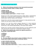
cuss the differential diagnosis in the light of radi- Squamous cell carcinoma is the most common
ological findings. histological type of bronchogenic carcinoma to
cavitate, followed by adenocarcinoma and large
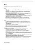
PATHOPHYSIOLOGY cell carcinoma14 (figure 2). Cavitation in a lung
Pathologically, a pulmonary cavity is defined as ‘a tumour is associated with poor prognosis with
gas-filled space within a zone of pulmonary conso- cavitation in squamous cell carcinoma being con-
lidation or within a mass or nodule, produced by the sidered a separate subentity related to worse
expulsion of a necrotic part of the lesion via the prognosis.15 Lymphomatoid granulomatosis,
bronchial tree’1 2 (figure 1). a rare Ebstein-Barr virus-driven B-cell lympho-
Cavity is also defined as an air-containing space proliferative disorder, can present with cavitary
within a pulmonary infiltrate or mass with a wall lung nodules due to necrosis. Pulmonary metas-
thicker than 4 mm.3 tases may also present as lung cavities, although
Many different pathological processes such as less frequently than primary lung malignancies
suppurative necrosis (pyogenic lung abscess), (figure 3). Cavitary metastases are mostly from
ischaemic necrosis (pulmonary infarction, malig- squamous cell carcinoma of head and neck ori-
nant tumour), caseous necrosis (tuberculosis) or gin, pelvic origin or oesophagal cancer.16 In
coagulative necrosis (radio-frequency ablation patients with HIV infection, rarely lymphoma
(RFA)) can cause a cavity. Pathogenic agents such and Kaposi sarcoma may develop cavitation.17
© Author(s) (or their as Mycobacterium tuberculosis, Klebsiella pneumo-
employer(s)) 2020. No niae and Staphylococcus aureus have a higher ten- Post-treatment appearance
commercial re-use. See dency to form cavitary lesions than other Primary or metastatic solid lung tumours may
rights and permissions. pathogens.4–6
Published by BMJ. develop cavities after chemotherapy.18 19 Central
necrosis and tumour cavitation following treatment
To cite: Canan A, Batra K, IMAGING MODALITIES with chemotherapeutic agents such as vascular
Saboo SS, et al. Postgrad Conventional chest radiography and computed endothelial growth factor inhibitors, which act by
Med J Epub ahead of print:
[please include Day Month tomography (CT) are the most commonly used ima- inhibition of angiogenesis, is indicative of good ther-
Year]. doi:10.1136/ ging modalities for evaluating pulmonary disease. apeutic response (figure 4). On the other hand, fill-
postgradmedj-2020- Although ultrasound could be useful for evaluation ing in of a cavity by regrowth of solid tumour may
138694 of pleural pathologies such as effusion or mass, it is indicate tumour progression.19
Canan A, et al. Postgrad Med J 2020;0:1–11. doi:10.1136/postgradmedj-2020-138694 1
, Review
Postgrad Med J: first published as 10.1136/postgradmedj-2020-138694 on 15 September 2020. Downloaded from http://pmj.bmj.com/ on September 19, 2020 at Karolinska BIBSAM
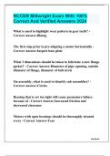
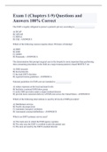
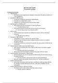
Figure 3 Cavitary metastasis of the lung secondary to (A) head and
neck cancer, (B) cervical cancer and (C) osteosarcoma.
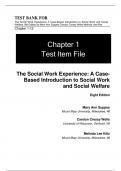
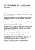
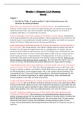
Figure 4 (A) Initial chest CT of a patient with non-small cell lung cancer
demonstrates a spiculated central soft tissue mass (arrow) in the right
lower lobe and multiple bilateral metastatic solid pulmonary nodules.
(B) Chest CT following immunotherapy with bevacizumab demonstrates
Consortia. Protected by copyright.
interval partial necrosis of the primary soft tissue lesion in the right lower
lobe (arrow) along with decrease in the size and density of the solid
pulmonary nodules suggesting response to treatment.
RFA is indicated in treatment of patients with inoperable pri-
mary or secondary lung tumours. Early post-ablation findings
include hyper-attenuating area larger than treated nodule due to
ensuing consolidation, inflammation and haemorrhage, which
progressively shrinks 2–3 weeks after treatment. Cavitation is
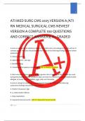
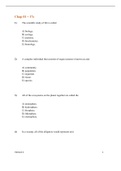
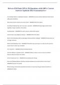
Figure 1 Schematic illustration of lung cavity. a common finding, occurring in up to 30% of the lesions between
1 and 3 months after RFA (figure 5). Development of cavitation is
accepted as a marker of treatment efficacy.20
Table 1 Major causes of pulmonary cavities Radiation-induced lung disease is common after radiation
C Cancer Bronchogenic carcinoma (most common SCC) therapy of lung tumours. Radiological findings include ground-
Metastasis (most common SCC) glass opacities or consolidation in the acute phase while trac-
A Autoimmune Granulomatosis with polyangiitis tion bronchiectasis, volume loss and scarring occur in the late
Rheumatoid arthritis phase (figure 6). Occasionally, there may be cavitation of the
V Vascular Pulmonary emboli lung parenchyma secondary to radiation-induced necrosis.
I Infection Tuberculosis Findings are usually limited to lung tissue within the radiation
Pulmonary abscess port.21
T Trauma Traumatic pulmonary pseudocyst
Y Youth Congenital pulmonary airway malformation
Pulmonary sequestration
Bronchogenic cyst
SCC, squamous cell carcinoma.
Figure 5 (A) Initial chest CT demonstrates a solid metastatic lesion in
the left lower lobe of the lung in a patient with the colon cancer. (B) Prone
intra procedure CT image shows intralesional ablation probe location
during radio-frequency ablation of the lesion. (C) Six-weeks follow-up
chest CT post-ablation shows development of a thin-walled relatively
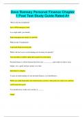
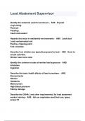
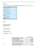
Figure 2 Cavitary lesions of the lung due to (A) primary squamous cell larger cavitary lesion in the location of prior solid nodule from coagulative
carcinoma, (B) primary adenocarcinoma and (C) lymphoma. The irregular or necrosis with surrounding larger ablation zone and adjacent localised
thickened wall is more often detected in malignant than benign lesions. parenchymal and pleural scarring suggesting response to treatment.
2 Canan A, et al. Postgrad Med J 2020;0:1–11. doi:10.1136/postgradmedj-2020-138694
Postgrad Med J: first published as 10.1136/postgradmedj-2020-138694 on 15 September 2020. Downloaded from http://pmj.bmj.com/ on September 19, 2020 at Karolinska BIBSAM
Radiological approach to cavitary lung lesions
Arzu Canan , Kiran Batra,1 Sachin S Saboo,2 Michael Landay,1 Asha Kandathil1
1
Department of Radiology, The ABSTRACT not helpful to evaluate parenchymal disease because
University of Texas Cavitary lesions in the lung are not an uncommon of the reverberation artefacts due to high air content
Southwestern Medical Center at
Dallas, Dallas, Texas, 75390, imaging encounter and carry a broad differential of the lungs.7 Despite recent developments, MRI
USA diagnosis that includes a wide range of pathological evaluation of the lung is still very limited due to
2
Department of Radiology, The conditions from cancers, infections/inflammatory low spatial resolution, high susceptibility and
University of Texas Health processes to traumatic and congenital lung abnormalities. motion artefacts.8 9 Although positron emission
Science Center at San Antonio,
San Antonio, Texas,
In this review article, we describe a comprehensive tomography is another modality, which is com-
78229, USA approach for evaluation of cavitary lung lesions and monly used to detect or characterise pulmonary
discuss the differential diagnosis in the light of nodules measuring >8 mm, it lacks specificity in
Correspondence to radiological findings. differentiating inflammatory from neoplastic
Arzu Canan, Department of lesions.10 Among all the imaging modalities, CT
Radiology, University of Texas remains the best and most sensitive technique.
Southwestern Medical Center
at Dallas, 5323 Harry Hines
Blvd., Dallas, TX, USA; arzu.
INTRODUCTION AETIOLOGY
A wide array of pathological conditions can present Cavitary lung lesions are caused by a wide variety of
as pulmonary cavities including cancers, infections, diseases such as malignancy, infection, inflammation
Received 15 July 2020 autoimmune (inflammatory) diseases, trauma and and congenital processes. The word ‘CAVITY’ can
Accepted 31 July 2020 congenital abnormalities.1 In view of the consider- be used as a mnemonic to remember and classify the
able phenotypic overlap that exists between the var- aetiology of a lung cavity (table 1).
Consortia. Protected by copyright.
ious lung cavitary conditions, a systematic approach
that requires cognitive recognition of likely possibi- C—cancer
lities, a grasp of morphological findings, compari- One of the most important steps in evaluation of
son to prior radiological exams and a search for a lung cavity is the differentiation between malig-
clinical clues will help to narrow the differential nant and benign causes. Primary lung cancer is
diagnosis. It is important to identify complications the leading cause of cancer-related death in men
as these may require appropriate management. The and women.11 The incidence of cavitation in pri-
aim of this review is to describe a comprehensive mary lung cancer detected by plain chest radio-
approach to evaluate cavitary lung lesions and dis- graphs and CT is 11%12 and 22%, respectively.13
cuss the differential diagnosis in the light of radi- Squamous cell carcinoma is the most common
ological findings. histological type of bronchogenic carcinoma to
cavitate, followed by adenocarcinoma and large
PATHOPHYSIOLOGY cell carcinoma14 (figure 2). Cavitation in a lung
Pathologically, a pulmonary cavity is defined as ‘a tumour is associated with poor prognosis with
gas-filled space within a zone of pulmonary conso- cavitation in squamous cell carcinoma being con-
lidation or within a mass or nodule, produced by the sidered a separate subentity related to worse
expulsion of a necrotic part of the lesion via the prognosis.15 Lymphomatoid granulomatosis,
bronchial tree’1 2 (figure 1). a rare Ebstein-Barr virus-driven B-cell lympho-
Cavity is also defined as an air-containing space proliferative disorder, can present with cavitary
within a pulmonary infiltrate or mass with a wall lung nodules due to necrosis. Pulmonary metas-
thicker than 4 mm.3 tases may also present as lung cavities, although
Many different pathological processes such as less frequently than primary lung malignancies
suppurative necrosis (pyogenic lung abscess), (figure 3). Cavitary metastases are mostly from
ischaemic necrosis (pulmonary infarction, malig- squamous cell carcinoma of head and neck ori-
nant tumour), caseous necrosis (tuberculosis) or gin, pelvic origin or oesophagal cancer.16 In
coagulative necrosis (radio-frequency ablation patients with HIV infection, rarely lymphoma
(RFA)) can cause a cavity. Pathogenic agents such and Kaposi sarcoma may develop cavitation.17
© Author(s) (or their as Mycobacterium tuberculosis, Klebsiella pneumo-
employer(s)) 2020. No niae and Staphylococcus aureus have a higher ten- Post-treatment appearance
commercial re-use. See dency to form cavitary lesions than other Primary or metastatic solid lung tumours may
rights and permissions. pathogens.4–6
Published by BMJ. develop cavities after chemotherapy.18 19 Central
necrosis and tumour cavitation following treatment
To cite: Canan A, Batra K, IMAGING MODALITIES with chemotherapeutic agents such as vascular
Saboo SS, et al. Postgrad Conventional chest radiography and computed endothelial growth factor inhibitors, which act by
Med J Epub ahead of print:
[please include Day Month tomography (CT) are the most commonly used ima- inhibition of angiogenesis, is indicative of good ther-
Year]. doi:10.1136/ ging modalities for evaluating pulmonary disease. apeutic response (figure 4). On the other hand, fill-
postgradmedj-2020- Although ultrasound could be useful for evaluation ing in of a cavity by regrowth of solid tumour may
138694 of pleural pathologies such as effusion or mass, it is indicate tumour progression.19
Canan A, et al. Postgrad Med J 2020;0:1–11. doi:10.1136/postgradmedj-2020-138694 1
, Review
Postgrad Med J: first published as 10.1136/postgradmedj-2020-138694 on 15 September 2020. Downloaded from http://pmj.bmj.com/ on September 19, 2020 at Karolinska BIBSAM
Figure 3 Cavitary metastasis of the lung secondary to (A) head and
neck cancer, (B) cervical cancer and (C) osteosarcoma.
Figure 4 (A) Initial chest CT of a patient with non-small cell lung cancer
demonstrates a spiculated central soft tissue mass (arrow) in the right
lower lobe and multiple bilateral metastatic solid pulmonary nodules.
(B) Chest CT following immunotherapy with bevacizumab demonstrates
Consortia. Protected by copyright.
interval partial necrosis of the primary soft tissue lesion in the right lower
lobe (arrow) along with decrease in the size and density of the solid
pulmonary nodules suggesting response to treatment.
RFA is indicated in treatment of patients with inoperable pri-
mary or secondary lung tumours. Early post-ablation findings
include hyper-attenuating area larger than treated nodule due to
ensuing consolidation, inflammation and haemorrhage, which
progressively shrinks 2–3 weeks after treatment. Cavitation is
Figure 1 Schematic illustration of lung cavity. a common finding, occurring in up to 30% of the lesions between
1 and 3 months after RFA (figure 5). Development of cavitation is
accepted as a marker of treatment efficacy.20
Table 1 Major causes of pulmonary cavities Radiation-induced lung disease is common after radiation
C Cancer Bronchogenic carcinoma (most common SCC) therapy of lung tumours. Radiological findings include ground-
Metastasis (most common SCC) glass opacities or consolidation in the acute phase while trac-
A Autoimmune Granulomatosis with polyangiitis tion bronchiectasis, volume loss and scarring occur in the late
Rheumatoid arthritis phase (figure 6). Occasionally, there may be cavitation of the
V Vascular Pulmonary emboli lung parenchyma secondary to radiation-induced necrosis.
I Infection Tuberculosis Findings are usually limited to lung tissue within the radiation
Pulmonary abscess port.21
T Trauma Traumatic pulmonary pseudocyst
Y Youth Congenital pulmonary airway malformation
Pulmonary sequestration
Bronchogenic cyst
SCC, squamous cell carcinoma.
Figure 5 (A) Initial chest CT demonstrates a solid metastatic lesion in
the left lower lobe of the lung in a patient with the colon cancer. (B) Prone
intra procedure CT image shows intralesional ablation probe location
during radio-frequency ablation of the lesion. (C) Six-weeks follow-up
chest CT post-ablation shows development of a thin-walled relatively
Figure 2 Cavitary lesions of the lung due to (A) primary squamous cell larger cavitary lesion in the location of prior solid nodule from coagulative
carcinoma, (B) primary adenocarcinoma and (C) lymphoma. The irregular or necrosis with surrounding larger ablation zone and adjacent localised
thickened wall is more often detected in malignant than benign lesions. parenchymal and pleural scarring suggesting response to treatment.
2 Canan A, et al. Postgrad Med J 2020;0:1–11. doi:10.1136/postgradmedj-2020-138694