
UCI BioSci D170, Williams Heart
Reading: Chapter 19
What are the two main functions of the heart?
The (right side of the) heart pumps oxygen-poor blood to the lungs via the pulmonary
circuit, and (left) heart pumps the oxygen-rich blood throughout the body via systemic
circuit.
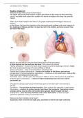
Where is the heart located in the thorax? Use proper anatomical terminology to discuss its
position.
In the thorax, the heart lies superior to the sternum/costal cartilage and rests superior to
the diaphragm. Oblique position: its apex lies left to the midline and more anterior than
the rest of the heart.
What is the pericardium? Describe the following layers of the pericardium:
A triple-layered sac that encloses the heart. From external to internal:
Fibrous pericardium – fused to the heart vessels superiorly and the diaphragm inferiorly. Holds
the heart in place and keeps it from overfilling w/ blood.
Serous pericardium – deep to the pericardium; double-layered.
Parietal layer of serous pericardium – fuses to the surface of the fibrous pericardium.
Visceral layer of serous pericardium (epicardium) – continuous w/ the parietal layer. Lies on the
heart and is considered the heart wall.
Pericardial cavity – slit-like space b/w the layers^. Filled w/ serous fluid produced by epithelium
lining the serous pericardium. -> reduces friction b/w beating heart and outer wall of
pericardial sac.
Describe the structure and function of the three layers of the heart wall. From external to
internal:
Epicardium – visceral layer of the pericardium. Often contains fat, especially in older people.
Myocardium – consists of cardiac muscle; heart layer that contracts. Connective tissues
arrange cardiac muscle cells into bundles that squeeze blood through the heart.
Endocardium – lines the heart chambers and covers their valves. A sheet of simple
squamous epithelium resting on connective tissue.
What are the four chambers of the heart?
Superiorly, there is the left and right atria, and below is the left and right ventricles.
, UCI BioSci D170, Williams Heart
Describe the structure and function of the internal chambers of the heart, noting the following
structures:
Interventricular septum – partition that divides the ventricles.
Interatrial septum – partition that divides the atria.
Coronary sulcus – horizontal groove that marks the boundary b/w the atria and ventricles.
Anterior interventricular sulcus – vertical groove that marks the anterior position of the
interventricular septum.
Posterior interventricular sulcus – groove that separates the ventricles on the heart’s
inferior (backside) surface.
Right atrium – receiving chamber for oxygen-poor blood returning from the body.
Superior and Inferior vena cava – veins that give blood to the atrium. Inferior enters
the chamber from behind.
Pectinate muscles – horizontal ridges that line the anterior part of the right atrium.
Crista terminalis – C-shaped ridge that separates the posterior and anterior right
atrium.
Right ventricle – receives blood from the right atrium and pumps it into the pulmonary
circuit. Atrium opens up through the tricuspid valve.
Pulmonary trunk – artery that receives the blood from the right ventricle.
Trabeculae carneae – irregular ridges of muscle that mark the ventricular walls.
Papillary muscles – cone-shaped projections that mark the ventricular walls.
Chordae tendineae – “heart strings.” Bands that project superiorly from the papillary
muscles to the tricuspid (atrioventricular) valve. Prevents closed cusps from flying
up. -> Don’t allow reflux of ventricular blood into the atria!
Left atrium – receiving chamber for oxygen-rich blood returning from the lungs.
Pulmonary veins – transfer blood from the lungs to the left atrium. 2 from the left
and 2 from the right.
Left ventricle – receives blood from the left atrium and pumps it into the systemic circuit.
Atrium opens up through the mitral valve. Has the same structures* as the right ventricle; forms
the apex of the heart. FUN FACT: It is the most muscular chamber (THICK myocardium).
Aorta – stem artery of the systemic circulation. Receives blood from the left ventricle via the
aortic semilunar valve.
Reading: Chapter 19
What are the two main functions of the heart?
The (right side of the) heart pumps oxygen-poor blood to the lungs via the pulmonary
circuit, and (left) heart pumps the oxygen-rich blood throughout the body via systemic
circuit.
Where is the heart located in the thorax? Use proper anatomical terminology to discuss its
position.
In the thorax, the heart lies superior to the sternum/costal cartilage and rests superior to
the diaphragm. Oblique position: its apex lies left to the midline and more anterior than
the rest of the heart.
What is the pericardium? Describe the following layers of the pericardium:
A triple-layered sac that encloses the heart. From external to internal:
Fibrous pericardium – fused to the heart vessels superiorly and the diaphragm inferiorly. Holds
the heart in place and keeps it from overfilling w/ blood.
Serous pericardium – deep to the pericardium; double-layered.
Parietal layer of serous pericardium – fuses to the surface of the fibrous pericardium.
Visceral layer of serous pericardium (epicardium) – continuous w/ the parietal layer. Lies on the
heart and is considered the heart wall.
Pericardial cavity – slit-like space b/w the layers^. Filled w/ serous fluid produced by epithelium
lining the serous pericardium. -> reduces friction b/w beating heart and outer wall of
pericardial sac.
Describe the structure and function of the three layers of the heart wall. From external to
internal:
Epicardium – visceral layer of the pericardium. Often contains fat, especially in older people.
Myocardium – consists of cardiac muscle; heart layer that contracts. Connective tissues
arrange cardiac muscle cells into bundles that squeeze blood through the heart.
Endocardium – lines the heart chambers and covers their valves. A sheet of simple
squamous epithelium resting on connective tissue.
What are the four chambers of the heart?
Superiorly, there is the left and right atria, and below is the left and right ventricles.
, UCI BioSci D170, Williams Heart
Describe the structure and function of the internal chambers of the heart, noting the following
structures:
Interventricular septum – partition that divides the ventricles.
Interatrial septum – partition that divides the atria.
Coronary sulcus – horizontal groove that marks the boundary b/w the atria and ventricles.
Anterior interventricular sulcus – vertical groove that marks the anterior position of the
interventricular septum.
Posterior interventricular sulcus – groove that separates the ventricles on the heart’s
inferior (backside) surface.
Right atrium – receiving chamber for oxygen-poor blood returning from the body.
Superior and Inferior vena cava – veins that give blood to the atrium. Inferior enters
the chamber from behind.
Pectinate muscles – horizontal ridges that line the anterior part of the right atrium.
Crista terminalis – C-shaped ridge that separates the posterior and anterior right
atrium.
Right ventricle – receives blood from the right atrium and pumps it into the pulmonary
circuit. Atrium opens up through the tricuspid valve.
Pulmonary trunk – artery that receives the blood from the right ventricle.
Trabeculae carneae – irregular ridges of muscle that mark the ventricular walls.
Papillary muscles – cone-shaped projections that mark the ventricular walls.
Chordae tendineae – “heart strings.” Bands that project superiorly from the papillary
muscles to the tricuspid (atrioventricular) valve. Prevents closed cusps from flying
up. -> Don’t allow reflux of ventricular blood into the atria!
Left atrium – receiving chamber for oxygen-rich blood returning from the lungs.
Pulmonary veins – transfer blood from the lungs to the left atrium. 2 from the left
and 2 from the right.
Left ventricle – receives blood from the left atrium and pumps it into the systemic circuit.
Atrium opens up through the mitral valve. Has the same structures* as the right ventricle; forms
the apex of the heart. FUN FACT: It is the most muscular chamber (THICK myocardium).
Aorta – stem artery of the systemic circulation. Receives blood from the left ventricle via the
aortic semilunar valve.


