
Oral Mucosa
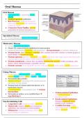
Oral Mucosa
• Oral epithelium + underlying lamina
propria
• Stratified squamous epithelium
• Rete Processes: epithelial extensions
projecting downwards into underlying
connective tissue
• Connective Tissue Papillae: upward
projections in between rete processes
Specialised Mucosa
• Covers dorsum of anterior 2/3 of tongue
Masticatory Mucosa
• Covers hard palate & gingiva
• Greater SA contact between epithelium & lamina propria
• Lamina propria forms continuum with periosteum – mucoperiosteum (no definite submucosa)
• Stratum basale: short columnar / cuboidal cells, contains stem cells, attaches to lamina propria at
basal lamina via hemidesmosomes
• Stratum spinosum (prickle): polygonal cells, larger than basal cells, larger nuclei than basal cells,
nuceloli appear, prickle spines visible (microscope artefact)
• Stratum granulosum: contains fine, basophilic keratohyaline granules within cytoplasm, cells
flatten, nuclei enlarge/elongate, organelles are lost
• Stratum corneum: most organelles are lost, cells are flattened with a tightly packed appearance
Lining Mucosa
• Covers buccal mucosal sulcus, labial mucosa,
ventral tongue surface, floor of mouth & soft palate Masticatory Lining
• No keratohyaline granules Basal Basal
• Submucosal layer consisting of fat, skeletal muscle Prickle Prickle
and minor salivary glands Granular Intermedium
• Stratum basale & stratum spinosum present, as in Cornified Superficial
masticatory mucosa
• No keratinisation, so no granular layer → stratum
intermedium
• Orthokeratinised Epithelium:
• No keratin on surface, so no cornified layer → cell nuclei disappear in
superficial layer keratinised layer
• Parakeratinised Epithelium:
Non-Keratinising Cells flattened, condensed nuclei
• Langerhan’s cells: found in stratum spinosum remain in cell cytoplasm
• Merkel cells: found in basal layer of gingival • Cornified Envelope: highly
epithelium, may function as touch receptors cross-linked layer of proteins in
• Melanocytes: melanin-producing cells in basal stratum corneum
layer of gingival epithelium
, Salivary Gland Physiology
Salivary Glands
• Exocrine; secrete onto mucosal surfaces of mouth
• Acinar cells: produce saliva, contain secretory granules
• Myoepithelial cells surround and stabilise acinar cells
• Parotid Gland: produces lots of amylase → serous secretions
• Submandibular Gland: produces amylase & mucous → seromucous secretions
• Sublingual Gland: produces lots of mucous → mucous secretion
• Minor Salivary Glands: present in most submucosal surfaces, secrete via main excretory
ducts of oral mucosa
Sympathetic stimuli
Afferent signals in sensory nerves
Salivary nuclei Thoracic spinal cord
Parasympathetic signals Sympathetic signals
Sublingual Parotid Submandibular
gland gland gland
Actions of Sympathetic NS
Formation of Primary Saliva • Causes most protein secretion
Parasympathetic: • Contracts myoepithelial cells
1. ACH binds to M3 receptor on acinar cell • Denervation → some gland
2. Intracellular [Ca] increases via inositol phosphate atrophy
mediators • Sends secretory signals to
3. Apical Cl channels open → Cl- moves out of cells submandibular and parotid
into lumen glands only
4. Na+ moves into lumen through tight junctions
down its gradient
5. H2O moves into lumen down osmotic gradient Actions of Parasympathetic NS
6. Na+, K+ and Cl- move into cell via a cotransporter • Causes most fluid secretion
Sympathetic:
• Contracts myoepithelial cells
1. NA binds to -adrenergic receptor on acinar cell
• Denervation → gland atrophy
2. Intracellular [cAMP] increases via Gs and
• Sends secretory signals all major
adenylyl cyclase
salivary glands
3. Exocytosis of proteins at apical membrane
• Vasodilation as part of secretory
4. NA also binds to -adrenergic receptors,
process
secreting small amounts of H2O and ions
Modification of Primary Saliva
• Resting/unstimulated (low flow rate) → striated ducts remove Na/Cl and secrete K
• Stimulated (high flow rate) → straited ducts remove Na/Cl and secrete K/HCO3-


