•
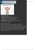
• Scrotum provides protection
• Epididymis is where spermatozoa is stored
• Vas deferens/sperm duct connects the epididymis and ejaculatory duct
• Testis are the glands that produce spermatozoa and hormones.
• Seminal vesicle produces many constituent ingredients of semen
• Prostate gland produces seminal fluid (the fluid that nourishes and transports sperm)
• Urethra allows urine to pass through the penis
The testes contain seminiferous tubules.
The cells that line the tubules undergo sperm formation and as they do, they move into the lumen.
At this point they are spermatozoa, and collect in the epididymis where they become motile.
They go to the vas deferens which carries sperm from the epididymis to the head of the penis during ejaculation.
While that happens, the seminal vesicles secrete mucus into the van deferens. Zinc-containing prostate fluid is
secreted in the prostate gland. These secretions are alkaline in order to: -maintain sperm mobility -provide
nutrients like sucrose, amino acids etc for energy -neutralise the acidity of leftover urine in the urethra
-neutralise the vaginal tract’s acidic pH
, • vagina is a canal that links the cervix of the uterus to the outside of the body. It accommodates the penis
during intercourse and is the birth canal
• Cervix opening from which the foetus exits the body
• Fallopian tubes connect the the uterus and the ovaries, the ovum travels down the tubes during ovulation
• Ovaries are where ova are produced and hormones are secreted.
• Uterus provides protection and supports the embryo/foetus up to birth
• Endometrium is the uterine lining. It thickens during the menstrual cycle and stays thick if implantation
happens to protect the foetus. It sheds (menstruation) in the absence of pregnancy.
Oocytes mature in the ovaries’ follicles. Follicles come from germinal epithelium around the ovaries. Matured
follicles go to the surface of the ovaries and a secondary follicle is released.
Cilia cells in the oviducal canal help the secondary oocyte to the oviduct.
The uterus wall has three linings.
1) perimetrium is the thin outside layer.
2) myometrium is the muscle layer
3) endometrium is a mucous membrane that has a good blood supply. It builds up/breaks down during the
menstrual cycle depending on whether or not implantation happens.
• Scrotum provides protection
• Epididymis is where spermatozoa is stored
• Vas deferens/sperm duct connects the epididymis and ejaculatory duct
• Testis are the glands that produce spermatozoa and hormones.
• Seminal vesicle produces many constituent ingredients of semen
• Prostate gland produces seminal fluid (the fluid that nourishes and transports sperm)
• Urethra allows urine to pass through the penis
The testes contain seminiferous tubules.
The cells that line the tubules undergo sperm formation and as they do, they move into the lumen.
At this point they are spermatozoa, and collect in the epididymis where they become motile.
They go to the vas deferens which carries sperm from the epididymis to the head of the penis during ejaculation.
While that happens, the seminal vesicles secrete mucus into the van deferens. Zinc-containing prostate fluid is
secreted in the prostate gland. These secretions are alkaline in order to: -maintain sperm mobility -provide
nutrients like sucrose, amino acids etc for energy -neutralise the acidity of leftover urine in the urethra
-neutralise the vaginal tract’s acidic pH
, • vagina is a canal that links the cervix of the uterus to the outside of the body. It accommodates the penis
during intercourse and is the birth canal
• Cervix opening from which the foetus exits the body
• Fallopian tubes connect the the uterus and the ovaries, the ovum travels down the tubes during ovulation
• Ovaries are where ova are produced and hormones are secreted.
• Uterus provides protection and supports the embryo/foetus up to birth
• Endometrium is the uterine lining. It thickens during the menstrual cycle and stays thick if implantation
happens to protect the foetus. It sheds (menstruation) in the absence of pregnancy.
Oocytes mature in the ovaries’ follicles. Follicles come from germinal epithelium around the ovaries. Matured
follicles go to the surface of the ovaries and a secondary follicle is released.
Cilia cells in the oviducal canal help the secondary oocyte to the oviduct.
The uterus wall has three linings.
1) perimetrium is the thin outside layer.
2) myometrium is the muscle layer
3) endometrium is a mucous membrane that has a good blood supply. It builds up/breaks down during the
menstrual cycle depending on whether or not implantation happens.