The Eye
Eye is the organ of vision - it converts light into electrical signals transmitted by neurons.
The shape of the eye is a slightly asymmetrical globe, whose diameter is about 25mm.
The visible front part of the eye consists of iris, cornea, pupil, sclera (the white part) and
conjunctiva (a clear layer of tissue covering the front of the eye, except the cornea).
The cornea
Is the front part of the eye.
It is a transparent layer (does not have blood vessels) with a thickness of about 0.6 mm.
Cornea represents the strongest part of the refracting power of the eye.
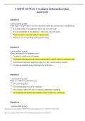
Fig: Internal parts of the eye
The iris
It is a thin, circular structure in the eye responsible for controlling the diameter and size of
the pupil and the amount of light reaching the retina.
The iris is a contractile structure, consisting mainly of smooth muscle surrounding the
pupil.
The pupil
Light enters the eye through the pupil, and the iris regulates the amount of light by
controlling the size of the pupil (similarly to the shutter in camera).
, Lens
Just behind the iris and pupil lies the lens.
The lens focuses light at the back of the eye.
Its about 9mm in diameter and 4 mm thick.
It is pliable, and changes shape for close focusing which is known as accommodation of
the eye.
Vitreous
Most of the eye is filled with a clear gel called the vitreous.
Retina
Light projects through the pupil and the lens to the back of the eye.
The inside lining of the eye is covered by special light-sensing cells that are collectively
called the retina.
Retina has photosensitive cells called rods and cones
Rods and cones convert light energy into electrical signals, that are carried to the brain by
the optic nerve.
Fovea centralis
In the middle of the retina is a small dimple called the fovea or fovea centralis.
This is the center of the eyes sharpest vision and the location of most color perception.
Optic nerve
The optic nerve is a bundle of nerve fibers that carries the electrical signal from the retina
to the brain for processing.
Eye is the organ of vision - it converts light into electrical signals transmitted by neurons.
The shape of the eye is a slightly asymmetrical globe, whose diameter is about 25mm.
The visible front part of the eye consists of iris, cornea, pupil, sclera (the white part) and
conjunctiva (a clear layer of tissue covering the front of the eye, except the cornea).
The cornea
Is the front part of the eye.
It is a transparent layer (does not have blood vessels) with a thickness of about 0.6 mm.
Cornea represents the strongest part of the refracting power of the eye.
Fig: Internal parts of the eye
The iris
It is a thin, circular structure in the eye responsible for controlling the diameter and size of
the pupil and the amount of light reaching the retina.
The iris is a contractile structure, consisting mainly of smooth muscle surrounding the
pupil.
The pupil
Light enters the eye through the pupil, and the iris regulates the amount of light by
controlling the size of the pupil (similarly to the shutter in camera).
, Lens
Just behind the iris and pupil lies the lens.
The lens focuses light at the back of the eye.
Its about 9mm in diameter and 4 mm thick.
It is pliable, and changes shape for close focusing which is known as accommodation of
the eye.
Vitreous
Most of the eye is filled with a clear gel called the vitreous.
Retina
Light projects through the pupil and the lens to the back of the eye.
The inside lining of the eye is covered by special light-sensing cells that are collectively
called the retina.
Retina has photosensitive cells called rods and cones
Rods and cones convert light energy into electrical signals, that are carried to the brain by
the optic nerve.
Fovea centralis
In the middle of the retina is a small dimple called the fovea or fovea centralis.
This is the center of the eyes sharpest vision and the location of most color perception.
Optic nerve
The optic nerve is a bundle of nerve fibers that carries the electrical signal from the retina
to the brain for processing.