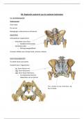
H5: Regionale anatomie van de onderste ledematen
5.1. De bekkengordel
Bekkengordel
Ossa coxae
Os sacrum
Bewegingen: anterversie en retroversie
Gewrichten
Articulationes cinguli pelvici
- Articulatio sacro-iliaca
o Nutatie/contranutatie
- Symphysis pubis
o Weinig bewegelijkheid
Vrouwen hebben een stompe hoek, mannen scherpe
Sacro-iliacaal gewricht
Tss beide facies auriculares
Versterkt door 3 ligamenten
- Lig. Sacro-iliacum ant.
- Ligg. Sacro-iliaca post.
- Ligg. Sacro-iliaca interossea
o Tss tuberositas ilium en
tuberositas van os sacrum
Proc. costales L5 naar crista iliaca = lig.
Ilium lumbale
,De symphysis pubis
discus pubis
verstevigd door 2 lig.
- Lig. pubicum inf.
- Lig. pubicum sup.
Extra ligamenten
Lig. sacrotuberale
- Ventrale zijde sacrum en deel ilium
NAAR tuber ischiadicum
Lig. sacrospinale
- Van sacrum & coccyx NAAR spina
ischiadica
4 doorgangen in OL
1. foramen ischiadicum majus
a. N. ischiadicus
b. N. gluteus inf. & sup.
2. Foramen ischiadicum minus
3. Doorgang onder lig. inguinale
a. A./V./N. femoralis
4. Canalis obturatorius 1
a. N. obturatorius
2
HERBEKIJK PLEXUS LUMBALIS & SACRALIS 3 4
+ bloedvaten van de OL (zie slides)
,5.2. Het heupgewricht – artrologie
Articulatio coxae
Kogelgewricht
- Caput femoris
o Lig. capitis femoris
- Acetabulum (facies lunata)
o Labrum acetabuli
o Lig. trans. acetabuli
Stabiel/stevig/gewrichtsdragend
Relatief beperkte mobiliteit
- Beendervormig
- Stevige ligamenten
Bewegingen
- Endorotatie/exorotatie
- Flexie/extensie
- Abductie/adductie
Flexiemogelijkheid beperkt door HAMSTRINGS
3 ligamenten beperken bewegingsmogelijkheid
1. Membrana synovialis (groen)
a. Smeert het gewricht
b. Tubulair rond. Lig. capitis femoris
c. Omslagplooi distaal collum
femoris
2. Membrana fibrosa (gewrichtskapsel)
a. Linea intertrochanterica vooraan
b. Rond synoviaal membraan
c. Proximaal v crista
intertrochanterica achteraan
, Ventraal zicht
Ventraal zicht
Lig. iliofemorale
- rond. Lig. capitis femoris
- Omslagplooi distaal collum femoris
- Beperkt extensie vd heup en retroversie
vh bekken Dorsaal zicht
Lig. pubofermorale
- Beperkt abductie vd heup
Lig. ischiofemoral
- Achterzijde acetabulum naar voor
Indeling spieren vd heup
Ventraal
- M. iliopsoas
Dorsaal: bilspieren:
- Dorsolaterale groep (oppervlakkig)
o M. gluteus maximus
o M. gluteus medius
o M. gluteus minimus
o M. tensor fasciae latae
- Pelvitrochanterische groep (diep)
o M. piriformis
o M. gemellus sup. & inf.
o M. obturatorius internus
o M. quadratus femoris
5.1. De bekkengordel
Bekkengordel
Ossa coxae
Os sacrum
Bewegingen: anterversie en retroversie
Gewrichten
Articulationes cinguli pelvici
- Articulatio sacro-iliaca
o Nutatie/contranutatie
- Symphysis pubis
o Weinig bewegelijkheid
Vrouwen hebben een stompe hoek, mannen scherpe
Sacro-iliacaal gewricht
Tss beide facies auriculares
Versterkt door 3 ligamenten
- Lig. Sacro-iliacum ant.
- Ligg. Sacro-iliaca post.
- Ligg. Sacro-iliaca interossea
o Tss tuberositas ilium en
tuberositas van os sacrum
Proc. costales L5 naar crista iliaca = lig.
Ilium lumbale
,De symphysis pubis
discus pubis
verstevigd door 2 lig.
- Lig. pubicum inf.
- Lig. pubicum sup.
Extra ligamenten
Lig. sacrotuberale
- Ventrale zijde sacrum en deel ilium
NAAR tuber ischiadicum
Lig. sacrospinale
- Van sacrum & coccyx NAAR spina
ischiadica
4 doorgangen in OL
1. foramen ischiadicum majus
a. N. ischiadicus
b. N. gluteus inf. & sup.
2. Foramen ischiadicum minus
3. Doorgang onder lig. inguinale
a. A./V./N. femoralis
4. Canalis obturatorius 1
a. N. obturatorius
2
HERBEKIJK PLEXUS LUMBALIS & SACRALIS 3 4
+ bloedvaten van de OL (zie slides)
,5.2. Het heupgewricht – artrologie
Articulatio coxae
Kogelgewricht
- Caput femoris
o Lig. capitis femoris
- Acetabulum (facies lunata)
o Labrum acetabuli
o Lig. trans. acetabuli
Stabiel/stevig/gewrichtsdragend
Relatief beperkte mobiliteit
- Beendervormig
- Stevige ligamenten
Bewegingen
- Endorotatie/exorotatie
- Flexie/extensie
- Abductie/adductie
Flexiemogelijkheid beperkt door HAMSTRINGS
3 ligamenten beperken bewegingsmogelijkheid
1. Membrana synovialis (groen)
a. Smeert het gewricht
b. Tubulair rond. Lig. capitis femoris
c. Omslagplooi distaal collum
femoris
2. Membrana fibrosa (gewrichtskapsel)
a. Linea intertrochanterica vooraan
b. Rond synoviaal membraan
c. Proximaal v crista
intertrochanterica achteraan
, Ventraal zicht
Ventraal zicht
Lig. iliofemorale
- rond. Lig. capitis femoris
- Omslagplooi distaal collum femoris
- Beperkt extensie vd heup en retroversie
vh bekken Dorsaal zicht
Lig. pubofermorale
- Beperkt abductie vd heup
Lig. ischiofemoral
- Achterzijde acetabulum naar voor
Indeling spieren vd heup
Ventraal
- M. iliopsoas
Dorsaal: bilspieren:
- Dorsolaterale groep (oppervlakkig)
o M. gluteus maximus
o M. gluteus medius
o M. gluteus minimus
o M. tensor fasciae latae
- Pelvitrochanterische groep (diep)
o M. piriformis
o M. gemellus sup. & inf.
o M. obturatorius internus
o M. quadratus femoris