
Summary Exam 2 Study Guide-Critical Care
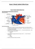
Exam 2 Study Guide-Critical Care Exam 2 Study Guide-Critical Care Basic Dysrhythmia Interpretation & Mgmt-Ch 7 Blood Flow Review Basic Electrophysiology • Automaticity-The ability for the heart muscle to generate it's own electrical activity o Pacemaker Cells arise naturally from the SA Node o Electrolytes involved are K+, Na+, & Ca+ Cardiac Action Potentials • Depolarization-occurs when charge is more (+) o P wave=atrial depolarization o QRS complex=ventricular depolarization o contraction o Systole • Repolarization-recharging period when charge is more (-) o T wave=ventricular repolarization o filling o Diastole • Electrical + Mechanical=Cardiac Contraction • Action Potential Curve o See Below-Sodium, Potassium, & Calcium flow in & out of the cell at different times allowing for charge changes and contraction of heart muscle tissue. o 4 Phases Early Repolarization • sodium channels close Phase 2-Plateau Phase • potassium leaves cell Phase 3-Rapid Repolarization • calcium channels close • potassium returns into cell quickly Phase 4-Resting Phase (polarized state) • active transport through the Na-K pump begins restoring K to inside the cell & sodium to the outside of the cell • cell membranes become impermeable to sodium • Potassium may move in & out of the cell Intrinsic Rates of Conduction Pathway • SA Node o natural pacemaker o 60-100 bpm • AV Node o takes over as pacemaker if SA node can't do the job o 40-60 bpm • Purkinje Fibers o last resort to pace heart if both SA & AV nodes fail o 20-40 bpm Cardiac Monitoring • 12 lead ECG o only 10 electrodes used to monitor, but gives 12 overall electrical pictures 4 limb leads 6 trunk leads o shows change or damage to heart muscle ischemia, infarct, enlarged cardiac chambers, electrolyte imbalances or drug toxicity Calculating Heart Rate from a 6 Second Strip • Graph Paper o Vertical boxes measure voltage or amplitude o Horizontal boxes measure time (in seconds) 1 small box=0.04 seconds 1 large box=0.20 seconds 5 large boxes=1 second • a six-second strip will be 30 large boxes • To calculate rate on a 6 second strip, count R-to-R and multiply by 10 Cardiac Waveforms & Determining Rhythm • P Wave • atrial depolarization • 0.04-0.10 seconds in duration Are they present? are they regularly occurring? is there a P for each QRS? are the P waves smooth, rounded, & upright? do all P waves look similar? • QRS Complex • ventricular depolarization • 0.06-0.10 seconds in duration is the complex 0.12 seconds (wide)? • can indicate ventricular origin is the complex 0.12 seconds (narrow)? • most likely supraventricular in origin (SA/AV nodes) do the complexes have a similar appearance across the tracing? • QT Interval • 0.38-0.42 seconds • begins at the QRS Complex to the end of the T wave • varies with heart rate • Pathological Q waves • 0.04 seconds in width & more than 1/4 R wave amplitude • indication of MI or myocardial tissue death • ST Segment • Elevation myocardial injury or hyperkalemia • STEMI looks like a tombstone • Depression myocardial ischemia or digoxin use • T Wave • ventricular repolarization • follows QRS Complex • U Wave • can sometimes bee seen after the T wave • can be normal or indicate hypokalemia • PR Interval • 0.12-0.20 seconds in duration is the interval 0.20 seconds? is the interval 0.12 seconds? is the interval constant across the tracing? Cardiac Dysrhythmias • Normal Sinus • Rate 60-100 • Rhythm regular • P waves before QRS & upright • PR interval 0.12-0.20 seconds • QRS complexes 0.12 seconds & look alike • Sinus tachycardia • Causes stress, exercise, fever, anemia, hypoxemia, CHF, pain • Effects on body faster rate decreases CO • S&S pounding heart, dizziness, anxiety, weak pulses decreased BP, UO • Treatment treat underlying cause give O2 if CHF, give Digoxin • Rate 100-160 • Rhythm regular • P waves before QRS & upright • PR interval 0.12-0.20 seconds • QRS complexes 0.12 seconds & look alike • Sinus bradycardia • Causes athletes, acute spinal cord injury • Effects on body lower rate decreases CO • S&S dizziness, SOB, weak pulses • Treatment Atropine by IV bolus to increase heart rate • 0.5 mg q3-5 minutes for a total dose of no more than 3 mg • S/Es: dry mouth/eyes, blurry vision, decreased UO, further slowing of heart rate if 0.5 mg are given • Rate 60 • Rhythm regular • P waves before QRS & upright • PR interval 0.12-0.20 seconds • QRS complexes 0.12 seconds & look alike Atrial Rhythms • Causes o Stress response, electrolyte imbalances, hypoxia, atrial injury, Dig toxicity, hypothermia o hyperthyroidism, alcohol, pericarditis, drugs • Premature Atrial Contractions (PACs) • early beats initiated by the atria not the SA node resulting in an early P wave • P waves & PR intervals may vary and are not at a regular rhythm • normal QRS Complexes • Precursor to other atrial tachycardias • S&S palpitations, heart skipping or pausing • Supraventricular tachycardia (SVT) • Causes heart disease, use of digoxin • Effects on body rhythm can come & go decreases CO • S&S • Treatment O2, start an IV, vagal maneuvers/valsalva, carotid massage adenosine by rapid IV push to block AV node transmission & stop heart briefly • 6 mg rapid IV push initially, followed by a second bolus of 12 mg 2 minutes later if not convert back to NSR o all doses followed by flush of NS and raising of arm • monitor HR, BP, and rhythm pattern • Have crash cart and/or defibrillator present in room • S/Es: diaphoresis, facial flushing, lightheadedness, chest pain Beta Blockers (Metroprolol, Labetalol) • decreases HR & BP, cardioprotective effects for MI patients • slow IV push • continuous monitoring of BP, HR, & rhythm o do NOT give for HR less than 50 or with 2nd or 3rd* heart block • S/Es: bradycardia, hypotension, heart failure Calcium Channel Blocker (Diltiazem, Verapamil) • slows conduction through AV node & decreases HR • Diltiazem 15-20 mg slow IV push over at least 2 minutes with a repeat bolus of 20-25mg if needed can be given after 15 minutes o IV infusion rate of 5-15 mg/hr may be started & titrated to desired HR • Verapamil 2.5-5 mg slow IV push over at least 2 minutes; if no response, can give repeat boluses of 5-10 mg every 15-30 minutes for a total dosage of 20 mg
Document information
- Uploaded on
- September 4, 2023
- Number of pages
- 44
- Written in
- 2023/2024
- Type
- Summary


