

Lecture 10: Basal Ganglia
The role of the Basal Ganglia in learning
The proposed function(s) of the basal ganglia are:
• selection of actions
• habit and skill acquisition
• goal-directed learning
Basal Ganglia Anatomy and Function
Basal ganglia circuitry (anatomical connections, neuronal subtypes, transmitter receptors etc.) is
highly conserved across vertebrate order e.g. mammals and lampreys share the same BG
organisation
Fundamental circuitry in the brain for controlling actions and for controlling learning
BG function therefore must be about basic processes conserved across species, occupying widely
different ecological niches and possessing different degrees of behavioural sophistication
BG Anatomy
The BG are made up by:
• Striatum - caudate and putamen in humans Commented [IM191]: The caudate and putamen in the human
is homologous to the striatum in rats
• Pallidum - globus pallidum, with internal and external compartments (GPi and GPe)
Commented [IM192]: Pallidum splits in to two different
• Substantia nigra - pars compacta and reticulata (SNc and SNr) functional parts
• Subthalamic nucleus (STN) Commented [IM193]: Source of dopamine to some parts of
the striatum and BG
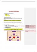
They represent a feedback and control circuit that are the interface between the neocortex and
the brainstem/midbrain areas that control and ‘release’ motor actions to create behaviours
Commented [IM194]: (Grillner and Robertson, 2016)
Not reference for this image, but for the entire anatomy and
circuitry section
,Striatum
• Majority of neocortical input; from the neocortical mantle and thalamus
o The thalamus(?) represents 45% of glutamatergic input to BG in the rat (Doug et al.
2010)
o Pallium is neocortical analogue for non-vertebrates
SNr/GPi
• Population of GABAergic tonically active (@ rest) neurons, thus constantly inhibiting motor
output
o Motor action controllers in brainstem/midbrains areas are constantly under
inhibitory control
o The BG circuits allow an action to take place by releasing the action from inhibition
• The SNr/GPi represents the output stations of the BG
• Their main outputs are:
o Thalamus (relays information back to cortex)
o Motor pattern generators in the midbrain and brainstem (controlling eye
movements, posture, swallowing, chewing, locomotion etc.)
• Importantly, 70% of the output axons from the SNr/GPi to motor centres have collaterals
reaching the thalamus and thus eventual the cortex
o Providing these structures with a copy of the motor commands issued
The SNr/GPi acts on Tectum/MLR/DLR (motor pattern generators/ motor action control centres) in
brainstem/midbrain areas to ‘release’ a motor command. This can occur via two control pathways
from the striatum; direct (D1/GO) or indirect (D2/STOP).
DIRECT PATHWAY (D1/GO)
Striatum —(inhibitory)—>SNr/GPi
• Inhibitory pathway - disinhibition of inhibitory SNr/GPi neurons
• GO pathway - releases motor programs from inhibition
Striatal medium spiny neurons (expressing D1 dopaminergic Rs and substance P) provide GABAergic
input to SNr/GPi, thus facilitating motor output
INDIRECT PATHWAY (D2/STOP)
Striatum—(inhibitory) à GPe—(inhibitory) à STN à SNr/GPi
• Two-step inhibition - inhibition of GPe neurons, thus reduced disinhibition of SNr/GPi, thus
motor pattern generators are inhibited and motor programmes not executed
• STOP - motor actions inhibited
Striatal medium spiny neurons (expressing D2 dopaminergic Rs and Enkephalin) provide GABAergic
input to GPe, which in turn provides GABAergic input to the STN. Striatal input thus activates STN,
which in turn provides excitation to SNr/GPi, producing a net inhibition of motor output
, HYPERDIRECT PATHWAY
• Directly excites the GPi capsule, thus inhibiting movement
• Frontal cortex (motor, cognitive, limbic) —> STN —> SNr/GPi
The STN also receives direct excitatory drive from the cortex and thalamus. Activation of the STN,
excites SNr/GPi and thus inhibits motor output
There is also dopamine in the BG circuitry that plays a very important role in controlling and
selecting actions
Dopamine [refs]
• SNc sends dopaminergic projections to striatum, which controls the function of the striatal
neurons and the ease with which motor programmes are ‘released’
o Also DA projections to motor control areas (Tectum/MLR/DLR) Commented [IM195]: Nigro-striatal dopaminergic pathway is
dysfunctional in PD due to degeneration à motor disabilities
BG BASICS
• Striatum forms the input, taking input from the neocortex
• SNr/GPi forms the output
• There are two primary projection pathways onto the GPi (output). These two pathways are
opposite and opposing in function:
o Allow action (DIRECT)
§ Striatum —(inhibitory)—>SNr/GPi
§ Disinhibition of output
o Inhibit action (INDIRECT)
§ Striatum—(inhibitory)—>GPe—(inhibitory)—->SNr/GPi
§ Inhibits the disinhibition of output
Action Selection and Production
The direct and indirect pathway can be separated at the level of the striatal neurons; by the genetic
profile of those striatal neurons.
• Striatal neurons that form a part of the direct projection —> internal capsule, have a genetic
profile (e.g. set of dopamine Rs) that are not shared with striatal neurons that instead
protect to the GP external capsule (forming the indirect pathway)
• We can separate the direct and indirect pathway by labelling the striatal neurons that lie
at the head of those to pathways
(Cui et al. 2013)
• D1 R - label direct pathway
• A2A R - label indirect pathway
o Used these genes to selectively label the direct and indirect pathways
What is the overall population activity in the striatal neurons contributing to either pathway when
the animal is performing an action?
• Task: animal responds to appropriate cue à press lever for food reward
o GCaMP - fluorescent reporter of [Ca] in neurons
The role of the Basal Ganglia in learning
The proposed function(s) of the basal ganglia are:
• selection of actions
• habit and skill acquisition
• goal-directed learning
Basal Ganglia Anatomy and Function
Basal ganglia circuitry (anatomical connections, neuronal subtypes, transmitter receptors etc.) is
highly conserved across vertebrate order e.g. mammals and lampreys share the same BG
organisation
Fundamental circuitry in the brain for controlling actions and for controlling learning
BG function therefore must be about basic processes conserved across species, occupying widely
different ecological niches and possessing different degrees of behavioural sophistication
BG Anatomy
The BG are made up by:
• Striatum - caudate and putamen in humans Commented [IM191]: The caudate and putamen in the human
is homologous to the striatum in rats
• Pallidum - globus pallidum, with internal and external compartments (GPi and GPe)
Commented [IM192]: Pallidum splits in to two different
• Substantia nigra - pars compacta and reticulata (SNc and SNr) functional parts
• Subthalamic nucleus (STN) Commented [IM193]: Source of dopamine to some parts of
the striatum and BG
They represent a feedback and control circuit that are the interface between the neocortex and
the brainstem/midbrain areas that control and ‘release’ motor actions to create behaviours
Commented [IM194]: (Grillner and Robertson, 2016)
Not reference for this image, but for the entire anatomy and
circuitry section
,Striatum
• Majority of neocortical input; from the neocortical mantle and thalamus
o The thalamus(?) represents 45% of glutamatergic input to BG in the rat (Doug et al.
2010)
o Pallium is neocortical analogue for non-vertebrates
SNr/GPi
• Population of GABAergic tonically active (@ rest) neurons, thus constantly inhibiting motor
output
o Motor action controllers in brainstem/midbrains areas are constantly under
inhibitory control
o The BG circuits allow an action to take place by releasing the action from inhibition
• The SNr/GPi represents the output stations of the BG
• Their main outputs are:
o Thalamus (relays information back to cortex)
o Motor pattern generators in the midbrain and brainstem (controlling eye
movements, posture, swallowing, chewing, locomotion etc.)
• Importantly, 70% of the output axons from the SNr/GPi to motor centres have collaterals
reaching the thalamus and thus eventual the cortex
o Providing these structures with a copy of the motor commands issued
The SNr/GPi acts on Tectum/MLR/DLR (motor pattern generators/ motor action control centres) in
brainstem/midbrain areas to ‘release’ a motor command. This can occur via two control pathways
from the striatum; direct (D1/GO) or indirect (D2/STOP).
DIRECT PATHWAY (D1/GO)
Striatum —(inhibitory)—>SNr/GPi
• Inhibitory pathway - disinhibition of inhibitory SNr/GPi neurons
• GO pathway - releases motor programs from inhibition
Striatal medium spiny neurons (expressing D1 dopaminergic Rs and substance P) provide GABAergic
input to SNr/GPi, thus facilitating motor output
INDIRECT PATHWAY (D2/STOP)
Striatum—(inhibitory) à GPe—(inhibitory) à STN à SNr/GPi
• Two-step inhibition - inhibition of GPe neurons, thus reduced disinhibition of SNr/GPi, thus
motor pattern generators are inhibited and motor programmes not executed
• STOP - motor actions inhibited
Striatal medium spiny neurons (expressing D2 dopaminergic Rs and Enkephalin) provide GABAergic
input to GPe, which in turn provides GABAergic input to the STN. Striatal input thus activates STN,
which in turn provides excitation to SNr/GPi, producing a net inhibition of motor output
, HYPERDIRECT PATHWAY
• Directly excites the GPi capsule, thus inhibiting movement
• Frontal cortex (motor, cognitive, limbic) —> STN —> SNr/GPi
The STN also receives direct excitatory drive from the cortex and thalamus. Activation of the STN,
excites SNr/GPi and thus inhibits motor output
There is also dopamine in the BG circuitry that plays a very important role in controlling and
selecting actions
Dopamine [refs]
• SNc sends dopaminergic projections to striatum, which controls the function of the striatal
neurons and the ease with which motor programmes are ‘released’
o Also DA projections to motor control areas (Tectum/MLR/DLR) Commented [IM195]: Nigro-striatal dopaminergic pathway is
dysfunctional in PD due to degeneration à motor disabilities
BG BASICS
• Striatum forms the input, taking input from the neocortex
• SNr/GPi forms the output
• There are two primary projection pathways onto the GPi (output). These two pathways are
opposite and opposing in function:
o Allow action (DIRECT)
§ Striatum —(inhibitory)—>SNr/GPi
§ Disinhibition of output
o Inhibit action (INDIRECT)
§ Striatum—(inhibitory)—>GPe—(inhibitory)—->SNr/GPi
§ Inhibits the disinhibition of output
Action Selection and Production
The direct and indirect pathway can be separated at the level of the striatal neurons; by the genetic
profile of those striatal neurons.
• Striatal neurons that form a part of the direct projection —> internal capsule, have a genetic
profile (e.g. set of dopamine Rs) that are not shared with striatal neurons that instead
protect to the GP external capsule (forming the indirect pathway)
• We can separate the direct and indirect pathway by labelling the striatal neurons that lie
at the head of those to pathways
(Cui et al. 2013)
• D1 R - label direct pathway
• A2A R - label indirect pathway
o Used these genes to selectively label the direct and indirect pathways
What is the overall population activity in the striatal neurons contributing to either pathway when
the animal is performing an action?
• Task: animal responds to appropriate cue à press lever for food reward
o GCaMP - fluorescent reporter of [Ca] in neurons


