OOGLEDEN
ANATOMIE & FYSIOLOGIE
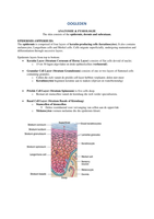
The skin consists of the epidermis, dermis and subcutaan.
EPIDERMIS (OPPERHUID)
The epidermis is comprised of four layers of keratin-producing cells (keratinocytes). It also contains
melanocytes, Langerhans cells and Merkel cells. Cells migrate superficially, undergoing maturation and
differentiation through succesive layers.
Epidermis layers from top to bottom:
Keratin Layer (Stratum Corneum of Horny Layer) consists of flat cells devoid of nuclei.
o 15 tot 30 lagen afgevlakte en dode epitheelcellen (verhoornd).
Granular Cell Layer (Stratum Granulosum) consists of one or two layers of flattened cells
containing granules.
o Cellen die zich vanuit de prickle cell layer hebben verplaatst; delen niet meer
o Keratinocyten beginnen keratine aan te maken (slijtvast en waterbestendig)
Prickle Cell Layer (Stratum Spinosum) is five cells deep.
o Bestaat uit stamcellen vanuit de kiemlaag die zich verder specialiseren.
Basal Cell Layer (Stratum Basale of Kiemlaag)
o Stamcellen of kiemcellen
Delen voortdurend voor vervanging van cellen aan de oppervlak
o Melanocyten vormen melanine die die epidermis kleurt.
,DERMIS (LEDERHUID OF CUTIS)
De dermis ligt onder epidermis en bevat een netwerk van bloedvaten, lymfevaten en zenuwvezels. De
dermis biedt mechanische sterkte, buigzaamheid en bescherming voor de ondergelegen weefsels.
Sebaceous Glands are located within eyebrow hairs.
Meibomian glands are modified sebacious glands found in the tarsal plates.
Consists of a central duct with multiple acini, the cells of which synthesize lipids (meibum)
that form the outer lipid layer of the tear film.
Glands of Zeiss
Modified sebaceous glands that secrete sebum
into the hair follicle, coating the eyelash shaft to
keep it from becoming brittle.
Glands of Moll
Modified apocriene sweat glands
o Secretion composed of parts of cellular
cytoplasm
Near the eyelash follicle
Duct empties into Zeiss gland or onto lid margin
between the lashes
Role in immune defense
Eccrine Sweat Glands
Distributed throughout the eyelid skin
Pilocebaceous Units
Comprise hair follicles together with their
sebaceous glands.
, NODULES & CYSTS
TERMINOLOGY
Vesicle. Circumscribed lesion containing serous fluid.
Bulla. Lare serous fluid-filled lesion.
Cyst. A nodule consisting of an epithelial-lined cavity filled with fluid or semi-solid material.
Plaque. Solid elevation of the skin, greater than 1 cm in diameter.
Papilloma. A benign neoplastic warty projections of the skin or mucous membrane.
Ulcer. A circumscribed area of epithelial loss.
, BENIGN LESIONS
CHALAZION (GRANULOMA)
Chalazion (Granuloma) is a meibomian cyst on the inside of the eyelid.
Signs.
o Chronische steriele granulomateuze ontsteking.
o Pijnloze nodule (soms astigmatisme) gevuld met lipogranuloma.
Etiology
o Verstopte Meibom klier of ander sebaceous klier.
o Sebum kan niet weg en hoopt op.
Treatment.
o A chalazion will often go away without treatment in a month or so.
o Warm Compress. Place a warm compress over the eyelid for 10 to 15 minutes at least
four times a day. Use lukewarm water.
o Surgery. If the chalazion continues to get bigger, it may need to be removed with
surgery.
o Steroid Injection (triamcinolone). May be preferred is chalazion is close to the
lacrimal punctum because of the risk of surgical damage.
o Systemische Tetracycline. May be required in patients with recurrent chalazia.
EPIDERMALE INCLUSIE CYST
Epidermal inclusion cyste is usually caused by implantation of epidermis into the dermis following
trauma or surgery.
Signs.
o Langzaam groeiend ronde, stevige, oppervlakkige of subcutane lesie
o Bevat keratine
Etiology.
o Cyste na trauma of operatie waarbij de epidermis de dermis in groeit.
,DERMOID CYST
Dermoid cyst is usualy subcutaneous or deeper and is typically attached to the periosteum at the lateral
end of the brow.
Subjective Signs.
o Subcutaan of dieper; keratiniseerd epitheel
o Congenitaal of eerste levensjaaren
o Verschillende plaatsen op lichaam
o Meer bij syndromen (Goldenhar)
o Meest voorkomende oculaire plaatsen
Superior temporale wenkbrauw
Buitenste canthus, onder ooglid (hoek)
Objective Signs.
o Vast stevig collageen oppervlak
o Gladde massa met huid erover
o Vast of minimaal beweegbaar
o Meest voorkomende orbitale lesie (ruimte
innemend proces zonder ontsteking) bij de
pediatrische populatie
o Anterior Lesies
Pijnloze massa op bovenooglid meestal bij laterale orbitale rand
Subcutaan, mobiele nodule, meestal frontozygomatic grens
o Diepe Lesie
Pijnloze progressieve diplopie
Proptosis, oogbewegings problemen, inferiore of superiore verplaatsing van
de globus.
Symptoms.
o Altijd pijnloos!
Etiologie.
o Onstaat tijdens embryonale ontwikkeling
SEBACEOUS (PILAR) CYST
Sebaceous (pilar) cyste is caused by a blocked pilosebaceous follicle and contains sebaceous
secretions.
Signs.
o Usually found on the eyelid; sometimes occurs at the inner canthus
,EPIDERMOID CYST
Epidermoid Cyste is uncommon and usually developmental, occurring along embryonic lines of
closure. Similar in appearance to epidermal inclusion cyste.
Mobiel subcutane op dermale massa
Ophoping (geschilferde cellen en keratine)
CYST OF ZEIS
Cyst of Zeis is a small, non-translucent cyt on the anterior lid
margin arising from obstructed glands of Zeis.
Signs
o Klein
o Niet doorschijnend
o Anteriore ooglidrand
Etiology
o Verstopte sebaceous klier
o Ass met wimper zakje
CYST OF MOLL (APOCRINE HIDROCYSTOMA)
Cyst of Moll is a small retention cyst of the lid margin
apocrine glands. It appears as a round, non-tender, translucent
fluid-filled lesion on the anterior lid margin.
Sings
o Kleine retention cyste
o Oolid rand
o Rond, niet gevoelig
o Doorzichtig
o Blauwige gloed
Etiology
o Verstopping apocrine klier
ECCRIENE HIDROCYSTOOM
Eccrine Hydrocystoom is less common, but similar in
appearance to a cyt of Moll, except that it is usually located
along the medial or lateral aspects of the lid, and is close to
but does not involve the lid margin itself.
Signs.
o Komt minder vaak voor
o Lijkt op cyste van Moll
o Mediale of laterale ooglid
o Dicht bij (maar niet op) de ooglid rand
o Retentie (ophoping) secretie
Etiology
o Obstructie eccrine zweet klieren
MILIA
Milia are caused by occlusion of pilosebaceous units
resulting in retention of keratin. They are tiny, white,
round, superficial papules that tend to occur in crops.
Signs.
o Retentie van keratine
o Kleine epidermaal cystes
o Witte en ronde oppervlakkige papules
o Vaak meerdere (crops)
Etiology.
o Occlusie van pilosebaceous units
,COMEDONES
Comedones are plugs of keratin and sebum within the dilated
orifice of hair follicales that often occur with patients with
acne.
Signs.
o Blackheads (open)
o Whiteheads (closed)
Etiology.
o Plugjes keratine of sebum
o Gedilateerde orifices van haarzakje
BENIGN EPIDERMAL TUMOURS
Papilloma is a benign neoplastic warty projection of the skin, mucous membrane
or glandular duct. A benign tumour derived from the epithelium.
Often seen with elderly
Aysmptomatic
Varied size and shape.
SQUAMOUS CELL (PLAVEISEL) PAPILLOMA
Squamous cell papilloma is a very common benign epithelial tumour with a variable clinical
appearance.
Signs
o Meest voorkomend benigne ooglid lesie
o Fingerlike projections of fibrovasculare connective tissue
covered by irregular squamous (plaveisel) epithelium
o Sessiele (brede basis) gesteelde lesie
Etiology
o Incidence increases with age
o Some cases result from virus infection
Behandeling
o Excision
, BASAL CELL PAPILLOMA
Basal cell papilloma is an extremely common slowly growing lesion found on the face, trunk and
extremities of elderly individuals.
Signs
o Meest voorkomend bij volwassenen (40-50)
o Afgebakende vettige bruine ‘plak’
o Brokkelig oppervlak (bloemkool)
o Vlak of geëleveerd; licht grijs of bruin
o Scherpe begrenzing
o Veranderd langzaam en veradelijk
o Komt voor op hele lichaam maar verhoogde kans op blootliggende huid
o Voorstadium kanker?
Etiology
o Groei van plaveisel epitheel
o Profliferatie van basaal cellen
Treatment
o Platte lesies afschaven
o Gesteelde lesies excisie
ANATOMIE & FYSIOLOGIE
The skin consists of the epidermis, dermis and subcutaan.
EPIDERMIS (OPPERHUID)
The epidermis is comprised of four layers of keratin-producing cells (keratinocytes). It also contains
melanocytes, Langerhans cells and Merkel cells. Cells migrate superficially, undergoing maturation and
differentiation through succesive layers.
Epidermis layers from top to bottom:
Keratin Layer (Stratum Corneum of Horny Layer) consists of flat cells devoid of nuclei.
o 15 tot 30 lagen afgevlakte en dode epitheelcellen (verhoornd).
Granular Cell Layer (Stratum Granulosum) consists of one or two layers of flattened cells
containing granules.
o Cellen die zich vanuit de prickle cell layer hebben verplaatst; delen niet meer
o Keratinocyten beginnen keratine aan te maken (slijtvast en waterbestendig)
Prickle Cell Layer (Stratum Spinosum) is five cells deep.
o Bestaat uit stamcellen vanuit de kiemlaag die zich verder specialiseren.
Basal Cell Layer (Stratum Basale of Kiemlaag)
o Stamcellen of kiemcellen
Delen voortdurend voor vervanging van cellen aan de oppervlak
o Melanocyten vormen melanine die die epidermis kleurt.
,DERMIS (LEDERHUID OF CUTIS)
De dermis ligt onder epidermis en bevat een netwerk van bloedvaten, lymfevaten en zenuwvezels. De
dermis biedt mechanische sterkte, buigzaamheid en bescherming voor de ondergelegen weefsels.
Sebaceous Glands are located within eyebrow hairs.
Meibomian glands are modified sebacious glands found in the tarsal plates.
Consists of a central duct with multiple acini, the cells of which synthesize lipids (meibum)
that form the outer lipid layer of the tear film.
Glands of Zeiss
Modified sebaceous glands that secrete sebum
into the hair follicle, coating the eyelash shaft to
keep it from becoming brittle.
Glands of Moll
Modified apocriene sweat glands
o Secretion composed of parts of cellular
cytoplasm
Near the eyelash follicle
Duct empties into Zeiss gland or onto lid margin
between the lashes
Role in immune defense
Eccrine Sweat Glands
Distributed throughout the eyelid skin
Pilocebaceous Units
Comprise hair follicles together with their
sebaceous glands.
, NODULES & CYSTS
TERMINOLOGY
Vesicle. Circumscribed lesion containing serous fluid.
Bulla. Lare serous fluid-filled lesion.
Cyst. A nodule consisting of an epithelial-lined cavity filled with fluid or semi-solid material.
Plaque. Solid elevation of the skin, greater than 1 cm in diameter.
Papilloma. A benign neoplastic warty projections of the skin or mucous membrane.
Ulcer. A circumscribed area of epithelial loss.
, BENIGN LESIONS
CHALAZION (GRANULOMA)
Chalazion (Granuloma) is a meibomian cyst on the inside of the eyelid.
Signs.
o Chronische steriele granulomateuze ontsteking.
o Pijnloze nodule (soms astigmatisme) gevuld met lipogranuloma.
Etiology
o Verstopte Meibom klier of ander sebaceous klier.
o Sebum kan niet weg en hoopt op.
Treatment.
o A chalazion will often go away without treatment in a month or so.
o Warm Compress. Place a warm compress over the eyelid for 10 to 15 minutes at least
four times a day. Use lukewarm water.
o Surgery. If the chalazion continues to get bigger, it may need to be removed with
surgery.
o Steroid Injection (triamcinolone). May be preferred is chalazion is close to the
lacrimal punctum because of the risk of surgical damage.
o Systemische Tetracycline. May be required in patients with recurrent chalazia.
EPIDERMALE INCLUSIE CYST
Epidermal inclusion cyste is usually caused by implantation of epidermis into the dermis following
trauma or surgery.
Signs.
o Langzaam groeiend ronde, stevige, oppervlakkige of subcutane lesie
o Bevat keratine
Etiology.
o Cyste na trauma of operatie waarbij de epidermis de dermis in groeit.
,DERMOID CYST
Dermoid cyst is usualy subcutaneous or deeper and is typically attached to the periosteum at the lateral
end of the brow.
Subjective Signs.
o Subcutaan of dieper; keratiniseerd epitheel
o Congenitaal of eerste levensjaaren
o Verschillende plaatsen op lichaam
o Meer bij syndromen (Goldenhar)
o Meest voorkomende oculaire plaatsen
Superior temporale wenkbrauw
Buitenste canthus, onder ooglid (hoek)
Objective Signs.
o Vast stevig collageen oppervlak
o Gladde massa met huid erover
o Vast of minimaal beweegbaar
o Meest voorkomende orbitale lesie (ruimte
innemend proces zonder ontsteking) bij de
pediatrische populatie
o Anterior Lesies
Pijnloze massa op bovenooglid meestal bij laterale orbitale rand
Subcutaan, mobiele nodule, meestal frontozygomatic grens
o Diepe Lesie
Pijnloze progressieve diplopie
Proptosis, oogbewegings problemen, inferiore of superiore verplaatsing van
de globus.
Symptoms.
o Altijd pijnloos!
Etiologie.
o Onstaat tijdens embryonale ontwikkeling
SEBACEOUS (PILAR) CYST
Sebaceous (pilar) cyste is caused by a blocked pilosebaceous follicle and contains sebaceous
secretions.
Signs.
o Usually found on the eyelid; sometimes occurs at the inner canthus
,EPIDERMOID CYST
Epidermoid Cyste is uncommon and usually developmental, occurring along embryonic lines of
closure. Similar in appearance to epidermal inclusion cyste.
Mobiel subcutane op dermale massa
Ophoping (geschilferde cellen en keratine)
CYST OF ZEIS
Cyst of Zeis is a small, non-translucent cyt on the anterior lid
margin arising from obstructed glands of Zeis.
Signs
o Klein
o Niet doorschijnend
o Anteriore ooglidrand
Etiology
o Verstopte sebaceous klier
o Ass met wimper zakje
CYST OF MOLL (APOCRINE HIDROCYSTOMA)
Cyst of Moll is a small retention cyst of the lid margin
apocrine glands. It appears as a round, non-tender, translucent
fluid-filled lesion on the anterior lid margin.
Sings
o Kleine retention cyste
o Oolid rand
o Rond, niet gevoelig
o Doorzichtig
o Blauwige gloed
Etiology
o Verstopping apocrine klier
ECCRIENE HIDROCYSTOOM
Eccrine Hydrocystoom is less common, but similar in
appearance to a cyt of Moll, except that it is usually located
along the medial or lateral aspects of the lid, and is close to
but does not involve the lid margin itself.
Signs.
o Komt minder vaak voor
o Lijkt op cyste van Moll
o Mediale of laterale ooglid
o Dicht bij (maar niet op) de ooglid rand
o Retentie (ophoping) secretie
Etiology
o Obstructie eccrine zweet klieren
MILIA
Milia are caused by occlusion of pilosebaceous units
resulting in retention of keratin. They are tiny, white,
round, superficial papules that tend to occur in crops.
Signs.
o Retentie van keratine
o Kleine epidermaal cystes
o Witte en ronde oppervlakkige papules
o Vaak meerdere (crops)
Etiology.
o Occlusie van pilosebaceous units
,COMEDONES
Comedones are plugs of keratin and sebum within the dilated
orifice of hair follicales that often occur with patients with
acne.
Signs.
o Blackheads (open)
o Whiteheads (closed)
Etiology.
o Plugjes keratine of sebum
o Gedilateerde orifices van haarzakje
BENIGN EPIDERMAL TUMOURS
Papilloma is a benign neoplastic warty projection of the skin, mucous membrane
or glandular duct. A benign tumour derived from the epithelium.
Often seen with elderly
Aysmptomatic
Varied size and shape.
SQUAMOUS CELL (PLAVEISEL) PAPILLOMA
Squamous cell papilloma is a very common benign epithelial tumour with a variable clinical
appearance.
Signs
o Meest voorkomend benigne ooglid lesie
o Fingerlike projections of fibrovasculare connective tissue
covered by irregular squamous (plaveisel) epithelium
o Sessiele (brede basis) gesteelde lesie
Etiology
o Incidence increases with age
o Some cases result from virus infection
Behandeling
o Excision
, BASAL CELL PAPILLOMA
Basal cell papilloma is an extremely common slowly growing lesion found on the face, trunk and
extremities of elderly individuals.
Signs
o Meest voorkomend bij volwassenen (40-50)
o Afgebakende vettige bruine ‘plak’
o Brokkelig oppervlak (bloemkool)
o Vlak of geëleveerd; licht grijs of bruin
o Scherpe begrenzing
o Veranderd langzaam en veradelijk
o Komt voor op hele lichaam maar verhoogde kans op blootliggende huid
o Voorstadium kanker?
Etiology
o Groei van plaveisel epitheel
o Profliferatie van basaal cellen
Treatment
o Platte lesies afschaven
o Gesteelde lesies excisie