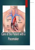
CARE OF THE PATIENT WITH A PEACE MAKER STUDY GUIDE
CARE OF THE PATIENT OBJECTIVES: Review normal conduction system of the heart Define Pacemaker List indications for the pacemaker Discuss pacemaker terminology Be able to use 3 letter code to discuss pacemaker Objectives Discuss types of malfunctions related to pacemakers Discuss patient assessment Discuss proper documentation Discuss indications and management of patient with an ICD Describe actions of magnet on pacemaker and ICD Symptomatic bradycardia A-V block Sick sinus syndrome Hypersensitive carotid sinus disease Conduction system trauma (accident or cardiac surgery) Chronic bifasicular or trifasicular block Indications Congenital anomalies Non reversible drug toxicities Junctional / Idioventricular Rhythm Overdrive Suppression of tachycardia’s Post operative Cardiac surgeries Low cardiac output state Firing: Pacemaker's generation of electrical stimuli Capture: Presence of a P or QRS or both after a pacemaker spike. Th Sensing: The pacemaker's ability to recognize the patient's own intrinsic rhythm Identification Code - 3 letters 1st - Chamber(s) paced ( A,V, Dual ) 2nd - Chamber(s) in which electrical activity is sensed ( A,V, D, O) 3rd - Response to sensing T Triggering of pacing function I - Inhibition of pacing function D - Dual response (i.e., any spontaneous atrial and ventricular activity will inhibit atrial and ventricular pacing, and no atrial activity will trigger a paced ventricular response) O - No response to an underlying electric signal (usually related to the absence of associated sensing function) Dual Chamber / DDD MODE: Two lead system with one lead wire in the atrium and one lead in the ventricle, capable of pacing the atria and ventricles, preserves AV synchrony. Single Chamber / VVI MODE: senses and paces the ventricle and is inhibited by a sensed ventricular event Single Chamber / AAI MODE:senses and paces in the atrium, and each sensed event triggers the generator to fire within the P wave NASPE/BPEG Generic Pacemaker Code Method of pacing Transcutaneous Transvenous Epicardial Permanent Transcutaneous Pacemaker External cardiac pacing is a temporary method of stimulating ventricular Myocardial depolarization through the chest wall via two large pacing Electrodes (patches). Temporary Transvenous Pacemaker: GOAL: Stimulate the myocardium to contract Established adequate cardiac output, blood pressure To ensure tissue perfusion to vital organs Administration of medication Emergency and elective clinical situations. Transvenous Pacing Pulse generator is externally attached Inserted through the vein(S/C, I/J, fem, Brachial) Single OR dual chamber Insulated wires with electrodes on the tip Wires Placed direct contact with endocardium ( Balloon ) Method of Transvenous pacing Pacing with PA catheterTemporary Atrial or ventricular pacing via thermoregulation PA catheter Assigned port for pacing wires Multiple capabilities Discrepancy in pacing while wedging ( PAOP ). Temporary Pacemaker NSA Transvenous Pacer Medtronic 5348 Single chamber temporary pace NSC Transvenous Pacer DUAL CHAMBER TEMPORARY EXTERNAL PACEMAKER Model 5392 NSF Transvenous Pacer Medtronic 5388 Dual chamber pacer Documentation for Pacemakers Patient assessment Type of pacing Patient underlying rhythm Pacemaker function Care of patient Permanent Pacemaker: Identify atrial activity Look for ventricular activity Assess patient’s hemodynamic status If pacemaker malfunctioning notify MD immediately Notify pacemaker company for interrogation as needed Lack of ventricular pacing is an emergency- ACLS protocol if indicated Care of Patient Transcutaneous/Temporary Pacing: Monitor VS hourly and prn Monitor patient level of comfort ECG strip document pacing function Evaluate pacemaker function, chart MA and rate Monitor patient’s underlying rhythm Evaluate patient HD response to pacing Troubleshoot as needed Change electrodes every 24 hours/prn Care of the Patient Transvenous Pacing: Monitor VS and HD response Evaluate ECG for presence of paced rhythm or resolution of initiating dysrhythmia Chart rate, type, sensing and capturing and the MA Monitor patient level of comfort Change dressing per policy Monitor for complications Monitor electrolytes Troubleshooting Failure to fire Pacemaker doesn’t fire when it should Troubleshooting cont… Failure to fire Reasons Electromagnetic interference Lead fracture Loose connections between the pulse generator and the lead wire Circuitry or battery failure Pacemaker turned off Treatment Move away from magnetic field Obtain a chest x-ray to determine lead position Reposition or replace lead if dislodged or fractured SQ pocket may need to be reopened to check connections Replace generator if needed Atropine and transcutaneous pacer at bedside Troubleshooting Cont.… Failure to sense: This occurs when the pacemaker fires too soon after an intrinsic beat and there are pacer spikes where there should not be. These can appear in the T wave, on the QRS or anywhere on the heart rhythm's tracing. Troubleshooting cont… Failure to sense Reasons Electromagnetic interference Electrolyte imbalances Lead fracture Loose connections between the pulse generator and the lead wire Battery failure Treatment Move away from magnetic field Obtain a chest x-ray to determine lead position Reposition or replace lead if dislodged or fractured May need to be reopened to check connections Look for the possible interference. Check electrolyte balance Troubleshooting cont… Failure to Capture: When loss of capture exists there is no P or QRS after the pacer has fired; just a spike. It is possible that pacing wire has lost contact with the chamber wall which can occur when the heart is too damaged to respond. Troubleshooting Continue… Failure to Capture: Reasons: Lead dislodgement Lead fracture Loose connections between pulse generator and lead wire Fibrosis at the tip of the lead causing changes in the pacing threshold/output Dead battery Treatment: Obtain a chest x-ray to determine lead position Reposition or replace lead if dislodged or fractured Have atropine and external pacemaker at bedside in case patient develops a symptomatic bradycardia Replace batteries OR replace pacemaker. Increased pacemaker output. Pacemaker Malfunctions Placement of pacing pads: Clinical Assessment of Patient with pacemaker Look for: Consciousness Blood pressure O2 saturation Symptoms Signs of inadequate perfusion Rhythm Strip Interpretation kind of pacemaker the patient has? Presence of an underlying rhythm? Presence of a pacer spike Presence of P waves (intrinsic or paced) and their rate and relationship to the QRS complexes Examine QRS complex If ventricular pacing, note whether each beat is preceded by a P wave (paced or intrinsic) Case Study: 63 year old female admitted to ICU with the c/o SOB secondary to pneumonia. She has positive history of CAD, CHF and PVD. Upon assessment, nurse find out that patient is more confused, lethargic and complaining of lightheadedness with cold and clammy skin. Patient monitor is now alarming with following vital signs HR 30 bpm palpable. BP: 80/40 RR: 14, SpO2: 92% @ 2 L N/C Pulse: Weak, palpable Q: If you are a bedside nurse for this patient what will be your intervention? Biventricular pacemaker Indicated for dilated cardiomyopathy with LBBB Both ventricles to contract simultaneously Increased quality of life Internal Cardiac Defibrillator Called ICD Used to prevent sudden cardiac death from VT/VF in cardiomyopathy and heart failure patients with EF less than or equal to 35 % Can be combined with dual chamber biventricular pacemaker in heart failure patients Life-Vest bridge to ICD CVP Placement in Patient with ICD During CVP insertion metal guidewire touching leads could trigger inappropriate shock Avoid metal guidewires Deactivate ICD during insertion Resuscitation of Patients With an ICD ACLS protocol should be initiated CPR should be initiated Anti-arrhythmics if indicated If external defibrillation required Can use magnet to deactivate CPR person could feel tingling sensation from patient if device shocks during CPR Place defibrillation pads 1 inch from device to avoid damage Anterior posterior pad placement when possible Medtronic “how to video” Model 5388
Document information
- Uploaded on
- July 10, 2023
- Number of pages
- 52
- Written in
- 2022/2023
- Type
- Exam (elaborations)
- Contains
- Questions & answers