

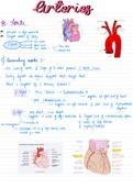
Arteries
* Aorta .
ventricle
situated
Left
•
in .
Largest vessels
of Body
-
.
Asc aorta
.
3
parts Aortic arch Thoracic Aorta
Desc .
aorta
Abdominal Hoda
1) Ascending aorta :
-
& valve
blue wall
of aorta
Cusps of its present 3HorkcSinus_
-
heart
During systole Hu
organs supplied
blood
Except
-
.
Heart is
supplied by 2
coronary Arteries : -
-
1) Right : -
Main Branch →
htow↳ᵈ '
heart
supplies Dwight part of .
post.inkwentiafwpge.is#
-
2. twentieth
bit
left ventricle & post part
.
of
septum .
2) Left : very
short 1cm
length ventricles
-
,
Supplies to walls
of Left &
Biggest part of interventricular septum
-
nt.sntuventn.at#cisumtbxBr
*
.
&
&
walls
Left Atrium
ofMuscles
papillary
"
↳ with be
of Right coronary artery
.
Angioma . .
vessels
Connection
of 2 .
,* Arteries Brain : 1)
of -
2)
Ant cerebral
Mid cerebral
A.
A.
4) Post
choroid
communicating
H
5) .
3) Ant
Communicating
"
*
wife : -
→
Hut Cerebral A.
.
→ Ant .
Communicating A.
→ Middle cerebral A.
->ost Communicating . A-
↳
Post Cerebral A.
Mpaa }
.
→ Basilar .
Vertebral A.→ Be Of subclavian A.
.
→ .
Post Cerebellar
→
.
Inf .
.
* Swbetaoian .
6th cervical vertebrae
Be .
Vertebral =
passes through Transverse foramen of
& enters cranial
cavity through foramen Magnum .
Inside skull Arteries
from Both side
joins & form Single Basilar
Artery
-
Gives Small Be Muscles Spinal cord & dusamakr 2
to
,
of
By
post cranial
fossa & 2
large
branches
Ant . spinal A.
. Past .
Spinal .
Gives
Dr. Thyiocervical If .
thyroid Tnf .
Laryngeal .
Hse .
Cervical .
Suprascapular .
Inf .
thoracic Musculophrenic A.
Sup Epigastric
.
* .
* Aorta .
ventricle
situated
Left
•
in .
Largest vessels
of Body
-
.
Asc aorta
.
3
parts Aortic arch Thoracic Aorta
Desc .
aorta
Abdominal Hoda
1) Ascending aorta :
-
& valve
blue wall
of aorta
Cusps of its present 3HorkcSinus_
-
heart
During systole Hu
organs supplied
blood
Except
-
.
Heart is
supplied by 2
coronary Arteries : -
-
1) Right : -
Main Branch →
htow↳ᵈ '
heart
supplies Dwight part of .
post.inkwentiafwpge.is#
-
2. twentieth
bit
left ventricle & post part
.
of
septum .
2) Left : very
short 1cm
length ventricles
-
,
Supplies to walls
of Left &
Biggest part of interventricular septum
-
nt.sntuventn.at#cisumtbxBr
*
.
&
&
walls
Left Atrium
ofMuscles
papillary
"
↳ with be
of Right coronary artery
.
Angioma . .
vessels
Connection
of 2 .
,* Arteries Brain : 1)
of -
2)
Ant cerebral
Mid cerebral
A.
A.
4) Post
choroid
communicating
H
5) .
3) Ant
Communicating
"
*
wife : -
→
Hut Cerebral A.
.
→ Ant .
Communicating A.
→ Middle cerebral A.
->ost Communicating . A-
↳
Post Cerebral A.
Mpaa }
.
→ Basilar .
Vertebral A.→ Be Of subclavian A.
.
→ .
Post Cerebellar
→
.
Inf .
.
* Swbetaoian .
6th cervical vertebrae
Be .
Vertebral =
passes through Transverse foramen of
& enters cranial
cavity through foramen Magnum .
Inside skull Arteries
from Both side
joins & form Single Basilar
Artery
-
Gives Small Be Muscles Spinal cord & dusamakr 2
to
,
of
By
post cranial
fossa & 2
large
branches
Ant . spinal A.
. Past .
Spinal .
Gives
Dr. Thyiocervical If .
thyroid Tnf .
Laryngeal .
Hse .
Cervical .
Suprascapular .
Inf .
thoracic Musculophrenic A.
Sup Epigastric
.
* .



