Chapter 17 : Cytoskeleton
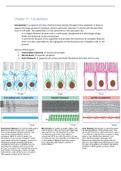
Cytoskeleton is a network of protein filaments that extends throughout the cytoplasm. It helps to
support the large volume of cytoplasm, which is particular important in animal cells because they
have no cell walls. The cytoskeleton is most prominent in the eukaryotic cell.
- It is a highly dynamic structure that is continuously reorganized as a cell changes shape,
divide and responses to the environment.
- It controls the location of the organelles and provides the machinery for transport between
them. It is also responsible for the segregation of chromosomes into 2 daughter cells at cell
division.
Consist of three parts:
1. Intermediate filaments → mechanical strength
2. Microtubules → organize cytoplasm
3. Actin filaments → supports cell surface and allows fibroblasts and other cell to crawl
,Intermediate filaments
- Main function: enable cells to withstand the mechanical stress that occurs when cells are
stretched.
- Found in: cytoplasm of most animal cells. They typically form a network throughout the
cytoplasm, surrounding the nucleus and extending out to the cell periphery → anchored to
desmosomes(where the plasma membrane is connected to that of another cell).
Also found in nucleus, called nuclear lamina. This strengthens nuclear envelope.
- Present in large number along nerve cell axons and muscle cells.
Characteristics:
- Like a rope where the strands of this cable are made of intermediate filament proteins.
Fibrous subunits each containing a central elongated rod domain with distinct unstructured
domains at the end.
- Interactions depend on noncovalent binding.
- The rod domains consist of alpha-helical rod domain(A) with unstructured regions at either
end. Pairs of monomers associate to form a dimer(B) and 2 dimers then line up to form an
antiparallel tetramer(C). Tetramers can pack together into a helical array containing 8
tetramer strands(D) which in turn assemble into the final ropelike intermediate filament(E)
,Intermediate filaments consist of 4 classes:
1. Keratin filaments (in epithelial cells)
2. Vimentin filaments (in connective-tissue cells)
3. Neurofilaments (in nerve cells)
4. Nuclear lamins (in nuclear envelope)
Keratin filaments
Are formed from a mixture of different keratin subunits. They typically span the interiors of epithelial
cells. The ends are connected to desmosomes.
Plectin is a accessory protein that reinforce and stabilize intermediate filaments → cross-link the
filaments into bundles and link them to microtubules, actin filaments and to adhesive structures in
the desmosomes. They are required to provide cells with the strength they need to withstand
mechanical stress.
Example: disease epidermolysis bullosa simplex cause the high vulnerability of the skin.
Nuclear envelope
Is constructed from a class of intermediate filament proteins called lamins. It disassembles and
reforms at each cell division, when the nuclear envelope breaks down during mitosis and then re-
forms in each daughter cell.
- The disassembly and reassembly of the nuclear lamina are controlled by the phosphorylation
and dephosphorylation of the lamins.
Example: disorder progeria cause affected individuals to age prematurely → children have wrinkled
skin, lose their teeth and hair.
Microtubules
Main function: part of the cytoskeleton and are mainly responsible for transporting and positioning
membrane-enclosed organelles within the cell and for guiding the intracellular transport →
organizing role in eukaryotic cells.
Found in: centrosome and cytoplasm.
, Characteristics:
- Microtubules are long hollow tubes of protein that can rapidly disassemble in 1 location and
reassemble in another.
- They grow out of the centrosome and create a system of tracks within the cell. Centrosomes
organizes an array of microtubules that radiates outward through the cytoplasm. The
centrosomes matrix consist of γ-tubulin which form a special type of tubulin.
- During cell division, cytoplasmic microtubules disassemble and then reassemble into the
mitotic spindle. This provides machinery that will segregate the chromosomes equally into
the 2 daughter cells.
- Can also form structures like cilia. These hairlike structures extend from the surface of many
eukaryotic cells, which use them either to swim or to sweep fluid over their surface.
- Dynamic instability = microtubule switches back and forth between polymerization and
depolymerization(growth) → allows them to undergo rapid remodeling; is crucial.
The centrosome can be compared to a fisherman casting a line: if there is no bite at the end
of the line, the line is quickly withdrawn, and a new cast is made; but, if a fish bites, the line
remains in place, tethering the fish to the fisherman.
Dynamic instability is driven by GTP hydrolysis. Each free tubulin dimer contains 1 GTP
molecule which is hydrolyzed to GDP shortly after the dimer is added to a growing
microtubule.
- Microtubule dynamics can be modified by drugs. They prevent the polymerization or
depolymerization of tubulin dimers → causes differences in behavior cell.
Example: colchicine ; cell stops in middle of mitosis because mitotic spindle disappears → kills
dividing cells.
- Cells are able to modify the dynamic instability of their microtubules for particular processes.
Example: cells specialized for secretion have their Golgi apparatus positioned toward the site
of secretion. The cell’s polarity is a reflection of the polarized systems of microtubules in its
interior, which help to position organelles in their required location within the cell and to
guide the streams of vesicular and macromolecular traffic moving between one part of the
cell and another. In the nerve cell, for example, all the microtubules in the axon point in the
same direction, with their plus ends toward the axon terminals; along these oriented tracks,
the cell is able to transport organelles, membrane vesicles, and macromolecules—either from
the cell body to the axon terminals or in the opposite direction
Structure and assembly of microtubules
Microtubules are built from subunits called tubulin. There is ∝-tubulin and 𝛽-tubulin, they are bound
tightly together by noncovalent bonding. This forms the wall of the hollow microtubule.
- Microtubules are polar, if they had no polarity they could not guide intracellular transport.
One end of the microtubule is thought to be the 𝛽-tubulin end which is called its + end and
the other one is ∝-tubulin and is called the – end.
Cytoskeleton is a network of protein filaments that extends throughout the cytoplasm. It helps to
support the large volume of cytoplasm, which is particular important in animal cells because they
have no cell walls. The cytoskeleton is most prominent in the eukaryotic cell.
- It is a highly dynamic structure that is continuously reorganized as a cell changes shape,
divide and responses to the environment.
- It controls the location of the organelles and provides the machinery for transport between
them. It is also responsible for the segregation of chromosomes into 2 daughter cells at cell
division.
Consist of three parts:
1. Intermediate filaments → mechanical strength
2. Microtubules → organize cytoplasm
3. Actin filaments → supports cell surface and allows fibroblasts and other cell to crawl
,Intermediate filaments
- Main function: enable cells to withstand the mechanical stress that occurs when cells are
stretched.
- Found in: cytoplasm of most animal cells. They typically form a network throughout the
cytoplasm, surrounding the nucleus and extending out to the cell periphery → anchored to
desmosomes(where the plasma membrane is connected to that of another cell).
Also found in nucleus, called nuclear lamina. This strengthens nuclear envelope.
- Present in large number along nerve cell axons and muscle cells.
Characteristics:
- Like a rope where the strands of this cable are made of intermediate filament proteins.
Fibrous subunits each containing a central elongated rod domain with distinct unstructured
domains at the end.
- Interactions depend on noncovalent binding.
- The rod domains consist of alpha-helical rod domain(A) with unstructured regions at either
end. Pairs of monomers associate to form a dimer(B) and 2 dimers then line up to form an
antiparallel tetramer(C). Tetramers can pack together into a helical array containing 8
tetramer strands(D) which in turn assemble into the final ropelike intermediate filament(E)
,Intermediate filaments consist of 4 classes:
1. Keratin filaments (in epithelial cells)
2. Vimentin filaments (in connective-tissue cells)
3. Neurofilaments (in nerve cells)
4. Nuclear lamins (in nuclear envelope)
Keratin filaments
Are formed from a mixture of different keratin subunits. They typically span the interiors of epithelial
cells. The ends are connected to desmosomes.
Plectin is a accessory protein that reinforce and stabilize intermediate filaments → cross-link the
filaments into bundles and link them to microtubules, actin filaments and to adhesive structures in
the desmosomes. They are required to provide cells with the strength they need to withstand
mechanical stress.
Example: disease epidermolysis bullosa simplex cause the high vulnerability of the skin.
Nuclear envelope
Is constructed from a class of intermediate filament proteins called lamins. It disassembles and
reforms at each cell division, when the nuclear envelope breaks down during mitosis and then re-
forms in each daughter cell.
- The disassembly and reassembly of the nuclear lamina are controlled by the phosphorylation
and dephosphorylation of the lamins.
Example: disorder progeria cause affected individuals to age prematurely → children have wrinkled
skin, lose their teeth and hair.
Microtubules
Main function: part of the cytoskeleton and are mainly responsible for transporting and positioning
membrane-enclosed organelles within the cell and for guiding the intracellular transport →
organizing role in eukaryotic cells.
Found in: centrosome and cytoplasm.
, Characteristics:
- Microtubules are long hollow tubes of protein that can rapidly disassemble in 1 location and
reassemble in another.
- They grow out of the centrosome and create a system of tracks within the cell. Centrosomes
organizes an array of microtubules that radiates outward through the cytoplasm. The
centrosomes matrix consist of γ-tubulin which form a special type of tubulin.
- During cell division, cytoplasmic microtubules disassemble and then reassemble into the
mitotic spindle. This provides machinery that will segregate the chromosomes equally into
the 2 daughter cells.
- Can also form structures like cilia. These hairlike structures extend from the surface of many
eukaryotic cells, which use them either to swim or to sweep fluid over their surface.
- Dynamic instability = microtubule switches back and forth between polymerization and
depolymerization(growth) → allows them to undergo rapid remodeling; is crucial.
The centrosome can be compared to a fisherman casting a line: if there is no bite at the end
of the line, the line is quickly withdrawn, and a new cast is made; but, if a fish bites, the line
remains in place, tethering the fish to the fisherman.
Dynamic instability is driven by GTP hydrolysis. Each free tubulin dimer contains 1 GTP
molecule which is hydrolyzed to GDP shortly after the dimer is added to a growing
microtubule.
- Microtubule dynamics can be modified by drugs. They prevent the polymerization or
depolymerization of tubulin dimers → causes differences in behavior cell.
Example: colchicine ; cell stops in middle of mitosis because mitotic spindle disappears → kills
dividing cells.
- Cells are able to modify the dynamic instability of their microtubules for particular processes.
Example: cells specialized for secretion have their Golgi apparatus positioned toward the site
of secretion. The cell’s polarity is a reflection of the polarized systems of microtubules in its
interior, which help to position organelles in their required location within the cell and to
guide the streams of vesicular and macromolecular traffic moving between one part of the
cell and another. In the nerve cell, for example, all the microtubules in the axon point in the
same direction, with their plus ends toward the axon terminals; along these oriented tracks,
the cell is able to transport organelles, membrane vesicles, and macromolecules—either from
the cell body to the axon terminals or in the opposite direction
Structure and assembly of microtubules
Microtubules are built from subunits called tubulin. There is ∝-tubulin and 𝛽-tubulin, they are bound
tightly together by noncovalent bonding. This forms the wall of the hollow microtubule.
- Microtubules are polar, if they had no polarity they could not guide intracellular transport.
One end of the microtubule is thought to be the 𝛽-tubulin end which is called its + end and
the other one is ∝-tubulin and is called the – end.