

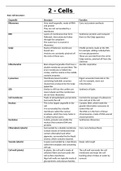
2 - Cells
Euk. Cell structure:
Organelle Structure Function
Ribosomes Very small organelle, made of RNA Carry out protein synthesis
and protein
They are not surrounded by a
membrane
RER Layers of membranes that form Synthesise proteins and transport
flattened, interconnected tubes them to the Golgi apparatus
through the cytoplasm
The outer face is covered in
ribosomes
Golgi Stacks of flattened, membrane- Modify proteins made at the rER,
bound sacs for example, adding carbohydrate
Vesicles are constantly pinched off to form glycoproteins
the ends of these sacs These are secreted from the cell in
Golgi vesicles, pinched off from the
cisternae
Mitochondria Bean-shaped organelles that have a Aerobic respiration
double membrane around them The
inner membrane is folded into
cristae, and the matrix in the middle
contains enzymes
Lysosomes Membrane-bound vesicles Digest unwanted materials in the
containing hydrolytic enzymes cell, for example, worn-out
(lysozymes) produced by the Golgi mitochondria
apparatus
SER Similar to rER but the cavities are Synthesis of lipids
more tubular and the membranes
do not have ribosomes
Cell membrane Made of phospholipids and proteins Controls the passage of substances
Surrounds the cell into and out of the cell
Nucleus This is the largest organelle in the Contains DNA, which holds the
cell genetic information necessary to
It is surrounded by a double control the cell
membrane called the nuclear The DNA is in the form of linear
envelope, which has many holes in chromosomes, made of DNA wound
it called nuclear pores around proteins
Nucleolus A dark, granular area inside the Synthesises ribosomes
nucleus that contains DNA and
proteins
Chloroplasts (plants) Surrounded by a double membrane Carry out photosynthesis
Contain stacks of membranes that
contain chlorophyll and other
pigments, surrounded by the liquid
stroma, which contains enzymes
Vacuole (plants) A space surrounded by a membrane Keeps cells turgid
called the tonoplast and containing
cell sap
Cell wall (plants) In plants, the cell wall is made of The cell wall surrounds the cell
cellulose fibres and surrounds the membrane and stops the cell
cell surface membrane bursting when it takes in water by
Algal cell walls are typically made of osmosis
glycoproteins and polysaccharides,
, and fungal cell walls are usually
made of the carbohydrate chitin
Prokaryotic cells:
- Have no nucleus/nuclear envelope
- Have single piece of DNA that is not associated w/ histones
- Don’t have membrane-bound organelles, e.g. mitochondria,
chloroplasts, ER, lysosomes or golgi
- Have cell wall made of murein
- Have ribosomes, but they’re smaller than in euk cells
- Contain plasmids -> small circular pieces of DNA tat carry DNA
additional to those in main piece of circular DNA
- Have flagella -> allows cell to move
- Has slimy capsule surrounding cell -> stops cell from drying out, helps protect cell from being engulfed by
WBC, and stores toxins
Viruses:
- Acellular -> non-living cells
- May contain DNA or RNA -> Surrounded by capsid, made of
many protein units
- Have attachment proteins on outside, which fits into a protein on
membrane of living host cell -> enables virus to attack cell
Microscopes:
Optical microscope:
- Works by passing a beam of light through a specimen, magnifying and focusing image using lenses
- Specimen needs to be thin, and may need to be stained to be seen more clearly
- Has a low resolution -> can’t clearly see 2 objects that are closer together than half the wavelength of light
being used in microscope = has longer wavelength
TEM:
- Uses a beam of e-, which has a smaller wavelength than light, so has a greater resolution than optical
microscope
- Must be a vacuum so that the e- beam can pass through
- Specimens have to be dead
- Produces 2D image
- Need to be aware of artefacts , e.g. dust
SEM:
- E- beam bounces of surface of object, allowing you to see the shape of structures like viruses
- Produces 3D image
- Specimen has to be thin before putting on a slide, however the structure can be altered by artefacts
Magnification:
Magnification = image size/actual size
Euk. Cell structure:
Organelle Structure Function
Ribosomes Very small organelle, made of RNA Carry out protein synthesis
and protein
They are not surrounded by a
membrane
RER Layers of membranes that form Synthesise proteins and transport
flattened, interconnected tubes them to the Golgi apparatus
through the cytoplasm
The outer face is covered in
ribosomes
Golgi Stacks of flattened, membrane- Modify proteins made at the rER,
bound sacs for example, adding carbohydrate
Vesicles are constantly pinched off to form glycoproteins
the ends of these sacs These are secreted from the cell in
Golgi vesicles, pinched off from the
cisternae
Mitochondria Bean-shaped organelles that have a Aerobic respiration
double membrane around them The
inner membrane is folded into
cristae, and the matrix in the middle
contains enzymes
Lysosomes Membrane-bound vesicles Digest unwanted materials in the
containing hydrolytic enzymes cell, for example, worn-out
(lysozymes) produced by the Golgi mitochondria
apparatus
SER Similar to rER but the cavities are Synthesis of lipids
more tubular and the membranes
do not have ribosomes
Cell membrane Made of phospholipids and proteins Controls the passage of substances
Surrounds the cell into and out of the cell
Nucleus This is the largest organelle in the Contains DNA, which holds the
cell genetic information necessary to
It is surrounded by a double control the cell
membrane called the nuclear The DNA is in the form of linear
envelope, which has many holes in chromosomes, made of DNA wound
it called nuclear pores around proteins
Nucleolus A dark, granular area inside the Synthesises ribosomes
nucleus that contains DNA and
proteins
Chloroplasts (plants) Surrounded by a double membrane Carry out photosynthesis
Contain stacks of membranes that
contain chlorophyll and other
pigments, surrounded by the liquid
stroma, which contains enzymes
Vacuole (plants) A space surrounded by a membrane Keeps cells turgid
called the tonoplast and containing
cell sap
Cell wall (plants) In plants, the cell wall is made of The cell wall surrounds the cell
cellulose fibres and surrounds the membrane and stops the cell
cell surface membrane bursting when it takes in water by
Algal cell walls are typically made of osmosis
glycoproteins and polysaccharides,
, and fungal cell walls are usually
made of the carbohydrate chitin
Prokaryotic cells:
- Have no nucleus/nuclear envelope
- Have single piece of DNA that is not associated w/ histones
- Don’t have membrane-bound organelles, e.g. mitochondria,
chloroplasts, ER, lysosomes or golgi
- Have cell wall made of murein
- Have ribosomes, but they’re smaller than in euk cells
- Contain plasmids -> small circular pieces of DNA tat carry DNA
additional to those in main piece of circular DNA
- Have flagella -> allows cell to move
- Has slimy capsule surrounding cell -> stops cell from drying out, helps protect cell from being engulfed by
WBC, and stores toxins
Viruses:
- Acellular -> non-living cells
- May contain DNA or RNA -> Surrounded by capsid, made of
many protein units
- Have attachment proteins on outside, which fits into a protein on
membrane of living host cell -> enables virus to attack cell
Microscopes:
Optical microscope:
- Works by passing a beam of light through a specimen, magnifying and focusing image using lenses
- Specimen needs to be thin, and may need to be stained to be seen more clearly
- Has a low resolution -> can’t clearly see 2 objects that are closer together than half the wavelength of light
being used in microscope = has longer wavelength
TEM:
- Uses a beam of e-, which has a smaller wavelength than light, so has a greater resolution than optical
microscope
- Must be a vacuum so that the e- beam can pass through
- Specimens have to be dead
- Produces 2D image
- Need to be aware of artefacts , e.g. dust
SEM:
- E- beam bounces of surface of object, allowing you to see the shape of structures like viruses
- Produces 3D image
- Specimen has to be thin before putting on a slide, however the structure can be altered by artefacts
Magnification:
Magnification = image size/actual size


